A dark spot beneath your fingernail can be alarming, particularly when you cannot recall any recent injury or trauma. These mysterious black dots, medically termed melanonychia, affect millions of people worldwide and present diagnostic challenges for both patients and healthcare professionals. While many cases prove benign and resolve naturally, others may signal serious underlying conditions requiring immediate medical intervention.
The appearance of nail pigmentation varies significantly in presentation, ranging from small isolated dots to extensive streaks extending across the entire nail plate. Understanding the various causes behind these discolourations becomes crucial for determining appropriate treatment pathways and identifying potentially life-threatening conditions such as subungual melanoma. Modern dermatological advances have revolutionised our ability to distinguish between harmless pigmentation and malignant transformations within the nail unit.
Contemporary research indicates that melanonychia affects approximately 77% of African Americans over age 20, while remaining relatively uncommon in Caucasian populations. This significant demographic variation highlights the importance of understanding both benign ethnic variants and pathological presentations when evaluating nail pigmentation disorders.
Subungual haematoma: Trauma-Induced melanonychia presentation
Subungual haematomas represent the most frequent cause of acute black nail discolouration, typically resulting from direct trauma to the nail bed. These blood collections beneath the nail plate create distinctive dark patches that evolve in colour and position over time. The initial appearance often presents as bright red or purple discolouration, gradually darkening to deep brown or black as the blood clots and begins to break down.
The pathophysiology involves rupture of small blood vessels within the highly vascularised nail bed, leading to extravasation of blood into the subungual space. This accumulation creates pressure beneath the nail plate, often causing significant pain and throbbing sensations. The trapped blood cannot escape through normal circulation, resulting in the characteristic persistent discolouration that moves distally with nail growth.
Acute nail bed haemorrhage following digital crush injuries
Crush injuries to the fingertips frequently result in immediate subungual haematoma formation, particularly when the trauma involves significant compressive forces. Common mechanisms include door entrapment, hammer strikes, heavy object impacts, and sports-related injuries. The severity of haemorrhage correlates directly with the magnitude of applied force and the duration of compression.
Clinical assessment requires evaluation of nail plate integrity, surrounding soft tissue damage, and potential underlying fractures. Digital crush injuries may compromise nail matrix function, leading to permanent nail deformities if not properly managed. Early intervention, including nail trephination for pressure relief, can prevent complications and preserve long-term nail aesthetics.
Chronic blood accumulation beneath nail plate keratin structure
Chronic subungual haematomas develop through repeated microtrauma or persistent pressure against the nail unit. This commonly occurs in athletes, particularly runners and tennis players, who experience repetitive toe impacts within poorly fitting footwear. The gradual accumulation of blood products creates persistent pigmentation that may persist for months.
Chronic presentations often lack the acute pain associated with sudden-onset haematomas, making them more challenging to diagnose accurately. The blood gradually organises and may calcify, creating irregular pigmentation patterns that can mimic more serious pathological processes. These cases require careful clinical correlation with patient history and activity patterns.
Distal subungual space bleeding patterns and colour variations
The location and distribution of subungual bleeding provide important diagnostic clues regarding the underlying mechanism and prognosis. Distal haematomas, located near the nail tip, typically result from direct trauma and resolve as the nail grows outward. Proximal bleeding patterns raise greater concern for nail matrix involvement and potential permanent sequelae.
Colour evolution follows predictable patterns, beginning with bright red acute bleeding, progressing through purple and brown phases, and eventually reaching deep black discolouration. The presence of multiple colour zones within a single haematoma indicates sequential bleeding episodes or varying degrees of blood degradation. Understanding these temporal changes helps clinicians estimate the age of injuries and plan appropriate interventions.
Post-traumatic nail matrix damage assessment criteria
Nail matrix evaluation following trauma requires systematic assessment of both immediate and delayed changes in nail production. The matrix, located beneath the proximal nail fold, generates the nail plate through continuous keratinocyte proliferation and differentiation. Trauma to this region can result in permanent nail dystrophies, including longitudinal ridging, splitting, and persistent pigmentation abnormalities.
Assessment criteria include nail plate thickness, surface texture, growth rate, and pigmentation patterns. Matrix scarring typically produces consistent abnormalities that persist throughout nail growth cycles, distinguishing permanent damage from temporary blood accumulation. Advanced imaging techniques, including high-frequency ultrasound, can visualise matrix architecture and predict long-term functional outcomes.
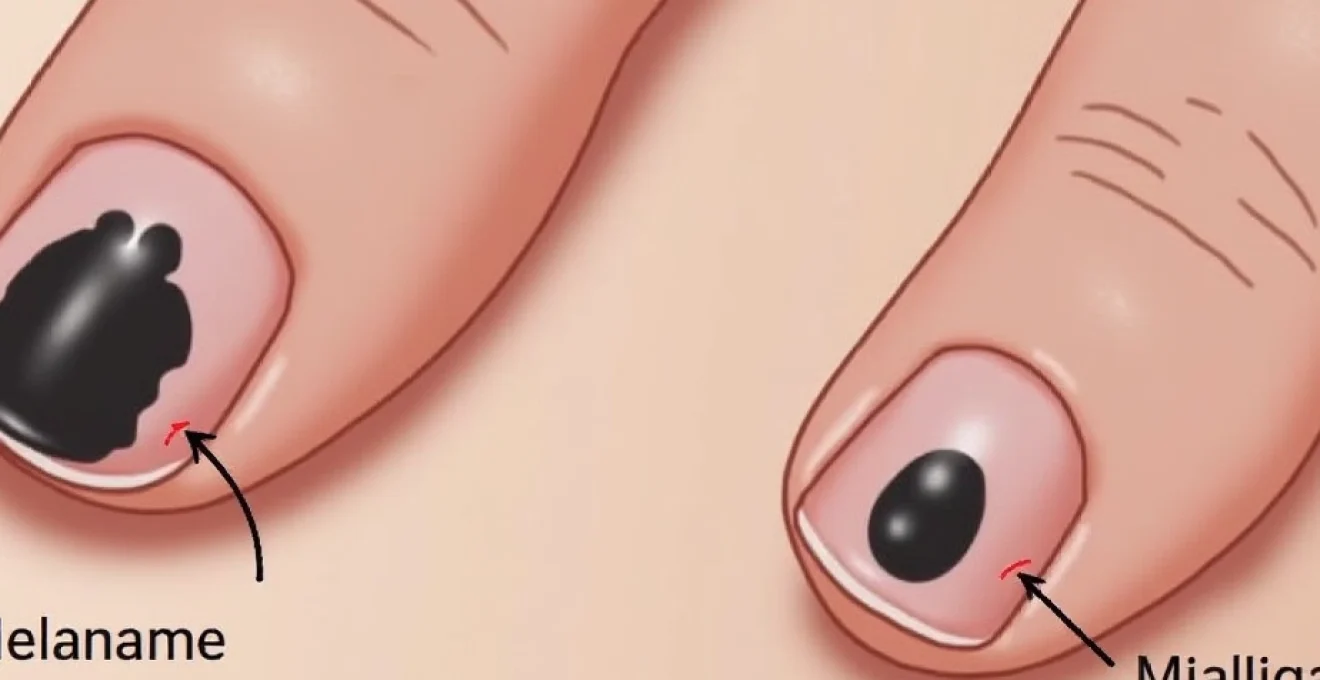
Malignant melanoma: subungual acral lentiginous melanoma detection
Subungual melanoma represents the most serious cause of nail pigmentation, accounting for 2-3% of all melanoma cases in Caucasian populations but comprising up to 25% of melanomas in individuals with darker skin types. This aggressive malignancy typically presents as expanding longitudinal pigmentation bands that may initially resemble benign melanonychia. Early detection becomes critical, as delayed diagnosis significantly worsens prognosis and survival rates.
The pathogenesis involves malignant transformation of melanocytes within the nail matrix or nail bed, leading to uncontrolled proliferation and melanin production. Unlike cutaneous melanoma, subungual variants rarely correlate with ultraviolet radiation exposure, instead arising through unknown mechanisms that may involve chronic mechanical trauma or genetic predisposition factors.
Recognition challenges arise from the rarity of this condition and its tendency to mimic benign processes. Many cases receive initial misdiagnosis as fungal infections, haematomas, or benign pigmentation, leading to delayed treatment and advanced disease stages at presentation. Healthcare providers must maintain high clinical suspicion when evaluating persistent or expanding nail pigmentation, particularly in high-risk demographic groups.
Hutchinson’s sign: periungual pigmentation extension diagnosis
Hutchinson’s sign represents a pathognomonic feature of advanced subungual melanoma, characterised by pigmentation extending beyond the nail plate onto the surrounding periungual skin. This finding indicates tumour invasion through the nail apparatus into adjacent cutaneous structures, typically signifying more advanced disease stages with increased metastatic potential.
The sign appears as brown or black discolouration involving the proximal nail fold, lateral nail folds, or hyponychium. Unlike normal ethnic pigmentation or benign melanonychia, Hutchinson’s sign demonstrates irregular borders, asymmetric distribution, and progressive expansion over time. The presence of this finding mandates immediate dermatological evaluation and tissue sampling for histopathological confirmation.
Early recognition of Hutchinson’s sign can significantly impact patient survival rates, as it often represents the transition from localised to invasive disease.
ABCDEF melanoma criteria applied to nail unit pathology
The traditional ABCDE melanoma criteria require modification for nail unit evaluation, leading to the development of the ABCDEF system specifically designed for subungual lesions. This adapted framework provides systematic assessment parameters for distinguishing malignant from benign nail pigmentation patterns.
The criteria include: Age (peak incidence 40-70 years), Band (brown-black colour with width ≥3mm), Change (rapid expansion or darkening), Diameter (involvement of thumb or great toe), Extension (Hutchinson’s sign), and Family history of melanoma. Combination assessment using multiple criteria provides enhanced diagnostic accuracy compared to individual parameter evaluation.
Amelanotic subungual melanoma atypical presentation forms
Amelanotic subungual melanomas lack characteristic pigmentation, presenting instead as pink, red, or flesh-coloured lesions that may resemble benign inflammatory conditions. These variants comprise approximately 20-30% of subungual melanoma cases and pose particular diagnostic challenges due to their atypical appearance and tendency to mimic other nail pathologies.
Clinical features include nail dystrophy, ulceration, bleeding, and nodular growth patterns. The absence of melanin deposition results from either decreased melanin production by malignant melanocytes or complete loss of melanocytic differentiation. Dermoscopy may reveal atypical vascular patterns, irregular surface architecture, and asymmetric growth characteristics that distinguish these lesions from benign inflammatory processes.
Nail matrix melanocyte transformation and pigment distribution
Malignant melanocyte transformation within the nail matrix creates distinctive pigmentation patterns that differ significantly from benign melanonychia. The nail matrix contains resident melanocytes that normally remain dormant but can undergo activation through various stimuli. Malignant transformation disrupts normal cellular regulation, leading to uncontrolled proliferation and irregular melanin distribution throughout the growing nail plate.
Histological examination reveals cellular atypia, increased mitotic activity, and loss of normal melanocyte organisation within the matrix. The resulting nail shows irregular pigmentation density, variable band width, and asymmetric colour distribution. Advanced cases demonstrate matrix destruction and replacement by malignant tissue, leading to nail dystrophy and eventual nail loss.
Breslow thickness measurement in acral melanoma cases
Breslow thickness measurement, the standard prognostic indicator for cutaneous melanoma, presents unique challenges in subungual locations due to anatomical constraints and tissue architecture differences. The measurement represents the vertical distance from the granular layer of the epidermis to the deepest point of tumour invasion, providing crucial staging information for treatment planning and prognosis determination.
Subungual measurements require careful histological orientation and recognition of tissue landmarks within the nail unit. The absence of typical epidermal architecture necessitates alternative reference points, including the nail plate-nail bed interface and underlying dermal structures. Accurate measurement becomes critical for determining appropriate surgical margins and adjuvant therapy requirements.
Benign melanonychia: ethnic and acquired pigmentation variants
Benign melanonychia encompasses a broad spectrum of nail pigmentation disorders that share similar clinical appearances but differ significantly in underlying pathophysiology and clinical significance. These conditions may arise from genetic factors, environmental exposures, inflammatory processes, or pharmacological interventions. Understanding the diverse presentations and causes becomes essential for appropriate clinical management and patient counselling.
The prevalence of benign melanonychia varies dramatically across ethnic groups, with individuals of African, Asian, and Hispanic descent showing significantly higher rates compared to Caucasian populations. This variation reflects differences in melanocyte activity, genetic susceptibility, and environmental factors that influence nail pigmentation patterns. Recognition of these ethnic variants prevents unnecessary invasive procedures and reduces patient anxiety regarding potential malignancy.
Racial melanonychia in fitzpatrick skin types IV-VI populations
Racial melanonychia represents a normal physiological variant in individuals with darker skin types, typically appearing as multiple longitudinal bands affecting several nails simultaneously. This condition results from increased baseline melanocyte activity within the nail matrix, leading to continuous melanin production and incorporation into the growing nail plate.
The bands typically appear symmetric, uniform in width and colour density, and remain stable over time. Multiple nail involvement constitutes a key distinguishing feature from malignant processes, which typically affect single nails with progressive changes. The condition may develop during childhood or adolescence and persists throughout life without requiring intervention.
Clinical recognition relies on understanding normal ethnic variation and avoiding misinterpretation as pathological processes. Patient education regarding the benign nature of racial melanonychia prevents anxiety and unnecessary medical interventions while establishing baseline documentation for future comparison.
Drug-induced melanonychia from antimalarial and chemotherapy agents
Numerous medications can induce nail pigmentation through various mechanisms, including direct melanocyte stimulation, inflammatory responses, and systemic metabolic effects. Antimalarial medications, particularly hydroxyeretamine and chloroquine, commonly cause diffuse nail darkening that affects multiple nails symmetrically. The pigmentation typically develops gradually over months of treatment and may persist for extended periods following drug discontinuation.
Chemotherapy-induced melanonychia affects up to 40% of patients receiving certain cytotoxic agents, including doxorubicin, bleomycin, and cyclophosphamide. The mechanism involves drug-induced inflammation within the nail matrix, leading to reactive melanocyte activation and increased melanin production. Recognition patterns include temporal correlation with treatment initiation, symmetric distribution, and gradual resolution following therapy completion.
Post-inflammatory hyperpigmentation following nail psoriasis
Inflammatory nail conditions, particularly psoriasis, can result in secondary hyperpigmentation through reactive melanocyte activation. Psoriatic nail involvement affects up to 90% of patients with psoriasis at some point during their disease course, creating various morphological changes including pitting, onycholysis, and discolouration patterns.
The inflammatory process triggers local cytokine release and growth factor production, stimulating dormant melanocytes within the nail matrix. This activation leads to increased melanin synthesis and incorporation into the nail plate, creating persistent pigmentation bands that may persist long after the inflammatory process resolves. The pigmentation typically correlates with disease severity and responds to effective psoriasis treatment.
Junctional naevi and nail matrix melanocytic proliferation
Junctional naevi within the nail matrix create benign melanonychia through localised melanocyte proliferation and increased melanin production. These benign proliferations typically remain stable over time and demonstrate consistent pigmentation patterns without the progressive changes characteristic of malignant processes.
Histological examination reveals organised melanocyte collections without cellular atypia or increased mitotic activity. The resulting nail pigmentation shows uniform density and sharp lateral borders, distinguishing these lesions from malignant counterparts. Long-term stability and absence of associated nail dystrophy provide additional reassuring clinical features that support benign diagnosis.
Fungal nail infections: onychomycosis chromatic manifestations
Fungal nail infections present diverse pigmentation patterns depending on the causative organism, infection location, and host immune response. While yellow discolouration represents the most common presentation, certain fungal species produce distinctive black or brown pigmentation that may be confused with other causes of nail darkening. Understanding these chromatic variations becomes essential for accurate diagnosis and appropriate antifungal therapy selection.
The pathophysiology involves fungal invasion of the nail plate structure, leading to keratin degradation and inflammatory responses. Some fungi produce pigmented compounds as byproducts of normal metabolism, while others create pigmentation through secondary inflammatory processes or interaction with host tissues. The location of infection influences pigmentation patterns, with proximal infections typically creating different colour distributions compared to distal involvement.
Certain dermatophyte species, including Trichophyton rubrum var. nigricans, can produce distinctive black pigmentation that closely mimics melanonychia or haematoma formation.
Diagnostic challenges arise when fungal infections coexist with other nail pathologies or when atypical presentations occur. Advanced diagnostic techniques, including potassium hydroxide preparation, fungal culture, and molecular testing, provide definitive identification of causative organisms and guide targeted therapy selection. Treatment response monitoring requires understanding of normal pigmentation evolution during successful antifungal therapy.
Bacterial nail pathology: pseudomonas and proteus discolouration
Bacterial nail infections create distinctive pigmentation patterns through direct bacterial pigment production and secondary inflammatory responses. Pseudomonas aeruginosa represents the most common cause of bacterial nail discolouration, producing characteristic blue-green pigmentation through pyocyanin production. However, certain Pseudomonas strains and other bacterial species can create black or dark brown discolouration that mimics other nail pathologies.
The infection typically occurs in moist environments where bacterial proliferation is enhanced, including chronic paronychia, nail trauma sites, or underlying nail dystrophy. The bacteria penetrate damaged nail structures and establish persistent colonisation that continues producing pigmented compounds throughout the infection course. Associated clinical features include nail softening, malodour, and surrounding tissue inflammation.
Proteus species can also cause dark nail discolouration through different metabolic pathways, typically producing brown to black pigmentation with associated nail dystrophy. These infections often occur secondary to trauma or immunocompromising conditions and may require systemic antibiotic therapy for resolution. Bacterial pigmentation typically shows rapid onset and progression, distinguishing it from slowly evolving melanonychia or chronic haematoma formation.
Clinical differential diagnosis using dermoscopy an
d nail biopsy techniques
Dermoscopy provides non-invasive magnified visualization of nail structures, enabling detailed assessment of pigmentation patterns and morphological features. The technique utilizes specialized instruments with 10-40x magnification and polarized lighting to reveal subsurface nail characteristics invisible to naked eye examination. Key dermoscopic features distinguishing bacterial infections include irregular pigment distribution, surface texture changes, and associated inflammatory signs around the nail fold.
Nail biopsy remains the gold standard for definitive diagnosis when clinical and dermoscopic findings prove inconclusive. The procedure involves sampling the nail matrix, nail bed, or nail plate depending on the suspected pathology location. Biopsy techniques include punch biopsy, excisional biopsy, and nail avulsion with matrix sampling. Proper specimen handling and histopathological interpretation require expertise in nail unit pathology to distinguish between benign and malignant processes.
Differential diagnosis considerations must account for patient age, medical history, nail involvement patterns, and associated clinical signs. Bacterial infections typically demonstrate rapid onset, asymmetric presentation, and response to antimicrobial therapy. The presence of purulent discharge, surrounding erythema, or systemic signs of infection supports bacterial etiology over other causes of nail pigmentation.
Advanced molecular diagnostic techniques, including polymerase chain reaction and mass spectrometry, provide rapid bacterial identification and antimicrobial susceptibility testing. These methods prove particularly valuable when traditional culture methods fail or when atypical organisms are suspected. Early accurate diagnosis enables targeted therapy selection and prevents progression to more serious complications including osteomyelitis or systemic bacterial dissemination.
Clinical correlation remains paramount in interpreting diagnostic test results, as laboratory findings must align with clinical presentation and patient risk factors. The integration of dermoscopic findings, histopathological results, and microbiological data provides comprehensive assessment capabilities that optimize diagnostic accuracy and treatment outcomes. Healthcare providers must maintain awareness of emerging bacterial pathogens and evolving antimicrobial resistance patterns that may influence both diagnostic approaches and therapeutic decisions.