The intersection of urinary function and sexual activity represents a complex physiological phenomenon that affects millions of women worldwide. Involuntary urination during intercourse, medically termed coital incontinence, occurs in approximately 24-60% of sexually active women who experience urinary incontinence. This condition stems from the intricate anatomical relationship between the female reproductive and urinary systems, where shared neural pathways, muscular structures, and hormonal influences create unique challenges during intimate moments. Understanding the underlying mechanisms, differentiating between various forms of fluid release, and exploring effective management strategies can significantly improve both physical comfort and psychological wellbeing for affected individuals.
Anatomical mechanisms of female urinary function during sexual activity
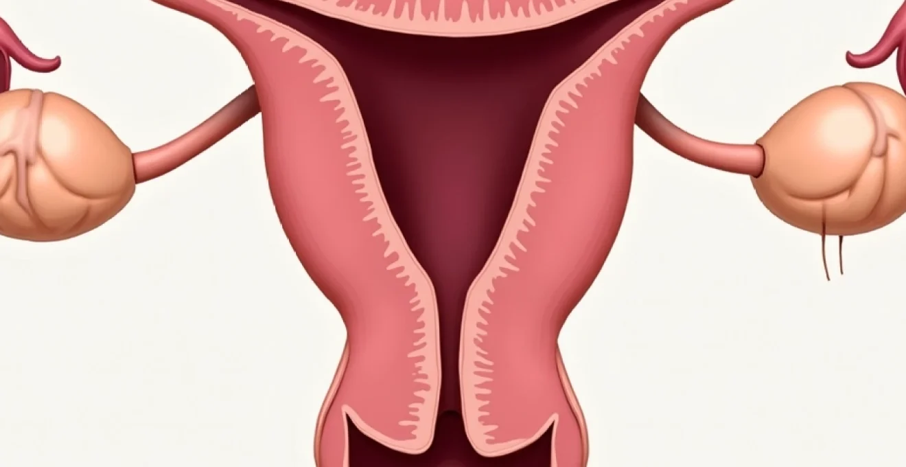
The female urogenital system demonstrates remarkable anatomical complexity, with the bladder, urethra, and reproductive organs sharing critical structural and functional elements. During sexual activity, these interconnected systems must coordinate their responses whilst maintaining continence under changing physiological conditions. The proximity of the bladder to the anterior vaginal wall means that mechanical pressure from penetration or movement can directly influence bladder function and urethral support mechanisms.
Urethral sphincter control and pelvic floor muscle coordination
The urethral sphincter complex consists of both internal smooth muscle and external striated muscle components that work in concert to maintain continence. The external urethral sphincter, composed of striated muscle fibres, is under voluntary control and forms part of the broader pelvic floor muscle group. During sexual arousal and activity, these muscles must maintain their supportive function whilst accommodating the physiological changes associated with increased pelvic blood flow and tissue engorgement.
Pelvic floor muscles, including the levator ani complex, provide crucial support to the urethrovesical junction—the critical anatomical point where the bladder meets the urethra. When these muscles are weakened through childbirth, ageing, or other factors, their ability to maintain urethral closure pressure during the increased intra-abdominal pressures of sexual activity becomes compromised. This muscular coordination becomes particularly challenging when the pelvic floor must simultaneously support continence whilst allowing for the relaxation necessary for comfortable penetration.
Bladder neck positioning changes during arousal and penetration
Sexual arousal triggers significant anatomical changes within the pelvis, including vascular engorgement and tissue swelling that can alter the normal positioning of urogenital structures. The bladder neck, which serves as a critical continence mechanism, may experience positional changes during penetrative activities. These alterations can affect the normal angle between the bladder and urethra, potentially compromising the natural anatomical barriers to urine leakage.
Research indicates that certain sexual positions may place greater mechanical stress on the bladder and urethral support structures. Positions that increase intra-abdominal pressure or directly compress the anterior vaginal wall can challenge even normally functioning continence mechanisms. The dynamic nature of sexual activity, with its varying pressures and movements, creates unique demands on the urethrovesical junction that differ significantly from the static pressures encountered during routine daily activities.
Neurological pathways: sympathetic and parasympathetic response interactions
The autonomic nervous system plays a fundamental role in both sexual response and bladder function, creating potential areas of conflict during intimate activity. Sympathetic nervous system activation, which occurs during sexual arousal, normally promotes bladder relaxation and urethral sphincter contraction—mechanisms that should theoretically support continence. However, the complex interplay between sexual excitement and bladder control can sometimes result in conflicting neural signals.
Parasympathetic stimulation, responsible for bladder contraction during normal voiding, can be inadvertently activated during sexual activity through various mechanisms. The shared neural pathways between genital sensation and bladder function mean that intense sexual stimulation may sometimes trigger inappropriate bladder contractions. This neurological crosstalk explains why some women experience urgency or actual leakage specifically during orgasmic responses.
Hormonal influences on detrusor muscle contractility during intercourse
Hormonal fluctuations significantly impact both sexual function and urinary continence, with oestrogen playing a particularly crucial role in maintaining urogenital tissue health. During sexual arousal, various hormones including oxytocin, prolactin, and endorphins are released, potentially influencing detrusor muscle behaviour. The detrusor muscle, which forms the main body of the bladder wall, may become hyperreactive under certain hormonal conditions, leading to involuntary contractions during sexual activity.
Oestrogen deficiency, common in postmenopausal women, leads to thinning and weakening of urogenital tissues, compromising natural continence mechanisms. This hormonal influence explains why coital incontinence often becomes more prevalent with advancing age, particularly following menopause when oestrogen levels decline significantly. The interplay between sexual arousal hormones and bladder function creates a complex physiological environment that can sometimes favour incontinence over normal continence mechanisms.
Physiological conditions that may cause involuntary urination during intercourse
Several distinct medical conditions can predispose women to experience involuntary urination during sexual activity. Understanding these underlying pathophysiological mechanisms is essential for developing targeted treatment approaches and helping affected individuals recognise that their symptoms represent treatable medical conditions rather than personal failings or unavoidable consequences of ageing.
Stress urinary incontinence: valsalva manoeuvre and intra-abdominal pressure
Stress urinary incontinence represents the most common underlying condition contributing to coital incontinence, affecting up to 37% of women at some point in their lives. This condition occurs when the normal support mechanisms for the urethra and bladder neck become compromised, typically through weakening of the pelvic floor muscles or damage to the connective tissue structures that maintain urethrovesical junction integrity.
During sexual activity, various movements and positions naturally increase intra-abdominal pressure through mechanisms similar to the Valsalva manoeuvre—the same physiological process that triggers stress incontinence during coughing, sneezing, or heavy lifting. The rhythmic movements of intercourse, combined with the muscular tension associated with sexual arousal and orgasm, create repetitive pressure spikes that can overwhelm weakened urethral support mechanisms. Women with pre-existing stress incontinence symptoms during daily activities are particularly vulnerable to experiencing leakage during sexual encounters.
Overactive bladder syndrome and detrusor hyperreflexia manifestations
Overactive bladder syndrome, characterised by involuntary detrusor muscle contractions, affects approximately 16% of women and can manifest dramatically during sexual activity. The intense physical and emotional stimulation of sexual encounters can trigger inappropriate bladder contractions in women with underlying detrusor hyperreflexia. These contractions may occur at any point during sexual activity but are particularly common during orgasmic responses when neural excitation reaches peak levels.
The phenomenon differs from stress incontinence in that it involves active bladder muscle contractions rather than passive leakage due to mechanical pressure. Women with overactive bladder may experience sudden, urgent sensations during sexual activity, sometimes accompanied by significant urine loss. This condition often coexists with daytime urgency symptoms, frequent urination, and nocturia, providing important diagnostic clues for healthcare providers.
Pelvic organ prolapse impact on urethrovesical junction stability
Pelvic organ prolapse, involving the descent of pelvic organs from their normal anatomical positions, significantly impacts urethrovesical junction stability and continence mechanisms. When the bladder, uterus, or rectum prolapse into the vaginal space, they can create mechanical interference with normal urethral support structures. This anatomical disruption becomes particularly problematic during sexual activity when additional mechanical forces are applied to already compromised support systems.
The degree of prolapse correlates with the severity of potential incontinence symptoms during intercourse. Even mild prolapse can create sufficient anatomical distortion to trigger leakage during the dynamic movements of sexual activity. The condition often develops gradually, meaning that women may notice increasing severity of coital incontinence over time as prolapse progresses. Comprehensive pelvic examination can identify prolapse and guide appropriate treatment strategies to address both the structural abnormality and its functional consequences.
Post-menopausal oestrogen deficiency and urogenital atrophy effects
The postmenopausal decline in oestrogen levels produces profound changes throughout the urogenital tract, collectively termed urogenital atrophy or genitourinary syndrome of menopause. These changes include thinning of urethral and bladder neck tissues, decreased collagen content in support structures, and reduced blood flow to pelvic tissues. The cumulative effect significantly compromises natural continence mechanisms and increases vulnerability to coital incontinence.
Oestrogen deficiency also affects vaginal tissues, leading to decreased lubrication and tissue elasticity that can make sexual activity uncomfortable and increase mechanical trauma to surrounding structures. The combination of weakened urethral tissues and potentially traumatic sexual experiences creates an environment particularly conducive to incontinence episodes. Localised oestrogen therapy can often dramatically improve these symptoms by restoring tissue health and supporting natural continence mechanisms.
Orgasmic incontinence: neurophysiological response patterns
Orgasmic incontinence represents a distinct phenomenon where urine leakage occurs specifically during climax, affecting approximately 15-20% of sexually active women. This condition differs from general coital incontinence in its timing and underlying neurophysiological mechanisms. During orgasm, the body experiences intense neural excitation that can sometimes overwhelm normal bladder control mechanisms, particularly in women with underlying continence vulnerabilities.
The orgasmic response involves complex interactions between the autonomic and somatic nervous systems, creating powerful muscular contractions throughout the pelvic region. These contractions, whilst normally coordinated to enhance sexual pleasure, can sometimes trigger inappropriate detrusor muscle activity in susceptible individuals. The phenomenon appears related to the intensity of orgasmic response, with more intense climaxes carrying higher risk for associated incontinence episodes.
Research suggests that orgasmic incontinence often correlates with overactive bladder symptoms in daily life, indicating shared underlying pathophysiology. Women experiencing this condition frequently report feeling betrayed by their bodies during moments of peak intimacy, highlighting the significant psychological impact beyond the physical symptoms. Understanding the neurophysiological basis helps normalise the experience and guides targeted treatment approaches.
The intense neural excitation during orgasm can create a perfect storm for incontinence in women with underlying bladder dysfunction, representing a treatable condition rather than an inevitable consequence of aging or childbirth.
Female ejaculation versus coital incontinence: clinical differentiation
Distinguishing between female ejaculation, squirting, and true coital incontinence represents a crucial clinical challenge with significant implications for treatment approaches and patient counselling. Female ejaculation involves the release of a small volume of thick, milky fluid from the Skene’s glands, located around the urethral opening. This fluid, analogous to male prostatic secretions, contains prostate-specific antigens and other proteins but typically involves volumes of only a few millilitres.
Squirting, conversely, involves the rapid expulsion of larger volumes of clear, watery fluid from the urethra during sexual stimulation. Recent research indicates that squirting fluid originates primarily from the bladder and consists largely of diluted urine mixed with prostatic-type secretions. The volumes involved can range from 30-150ml, significantly more than true female ejaculation but representing normal physiological responses to intense sexual stimulation rather than pathological incontinence.
True coital incontinence differs from both ejaculation and squirting in several key characteristics. The fluid is typically yellow in colour with the characteristic odour of concentrated urine, and the volume can range from a few drops to complete bladder emptying. Importantly, coital incontinence is involuntary and often distressing to the individual, occurring independently of sexual arousal levels or orgasmic response. The timing may be unpredictable, happening during penetration, movement, or at any point during sexual activity.
Clinical evaluation should focus on fluid characteristics, timing of occurrence, associated symptoms, and the individual’s subjective experience. Women experiencing true coital incontinence often report embarrassment, avoidance of sexual activity, and significant impact on relationship quality. This psychosocial dimension helps differentiate pathological incontinence from normal physiological responses that, whilst potentially surprising, do not typically cause ongoing distress or functional impairment.
| Characteristic | Female Ejaculation | Squirting | Coital Incontinence |
|---|---|---|---|
| Volume | Small (few millilitres) | Moderate to large (30-150ml) | Variable (drops to full bladder) |
| Colour | Whitish/milky | Clear to slightly yellow | Yellow |
| Odour | Mild, non-urine | Minimal to slight urine | Strong urine odour |
| Timing | During orgasm | During intense stimulation | Unpredictable during activity |
| Psychological Impact | Generally neutral to positive | Variable, often neutral | Typically distressing |
Preventive interventions and pelvic floor rehabilitation techniques
Effective management of coital incontinence relies heavily on comprehensive pelvic floor rehabilitation programmes that address the underlying muscular and neurological deficits contributing to symptoms. The approach must be individualised based on the specific type of incontinence, severity of symptoms, and contributing factors identified through clinical assessment. Success rates for conservative management approaches range from 60-85%, making these interventions the first-line treatment for most women experiencing coital incontinence.
Kegel exercise protocols for urethral sphincter strengthening
Pelvic floor muscle training, commonly known as Kegel exercises, forms the cornerstone of conservative treatment for coital incontinence. However, proper technique is crucial for effectiveness, as studies indicate that up to 50% of women perform these exercises incorrectly without professional guidance. The optimal protocol involves both slow-twitch and fast-twitch muscle fibre training to address different aspects of continence control.
Slow-twitch training involves sustained contractions held for 10 seconds, repeated 10 times, three times daily. These exercises build the endurance capacity necessary for maintaining continence during prolonged activities. Fast-twitch training incorporates rapid contractions performed in sets of 10, designed to improve the quick reflexive responses needed during sudden pressure increases, such as those occurring during sexual activity.
Progressive overload principles should be applied, gradually increasing contraction duration and intensity over 8-12 weeks. Many women benefit from combining pelvic floor contractions with functional activities that simulate the demands of sexual positions. For example, performing Kegel exercises whilst in various sexual positions can help develop the specific muscle memory and strength patterns needed for real-world continence maintenance.
Biofeedback training methods for pelvic floor muscle coordination
Biofeedback training significantly enhances the effectiveness of pelvic floor rehabilitation by providing real-time information about muscle activity patterns. Surface electromyography (sEMG) sensors placed around the pelvic floor muscles allow women to visualise their muscle contractions, ensuring proper technique and identifying compensatory patterns that may compromise treatment success.
The training typically involves 6-8 sessions over 8-12 weeks, during which women learn to isolate pelvic floor muscles from accessory muscle groups such as the gluteals and abdominals. Proper muscle isolation is essential because compensatory patterns can actually worsen incontinence by increasing intra-abdominal pressure whilst failing to strengthen the target muscles. Advanced biofeedback protocols incorporate pressure sensors that help women learn to coordinate pelvic floor contractions with breathing patterns and functional movements.
Home biofeedback devices are now available, allowing women to continue training between clinical sessions. These devices provide immediate feedback about contraction strength and duration, helping maintain motivation and ensuring continued progress. Success rates for biofeedback-assisted pelvic floor training reach 70-80% for stress incontinence symptoms,
with many women reporting significant improvement in symptoms when consistent training protocols are followed.
Electrical stimulation therapy applications in incontinence management
Electrical stimulation therapy offers a valuable adjunct to traditional pelvic floor training, particularly for women who struggle to achieve effective voluntary muscle contractions. Low-frequency electrical stimulation (typically 10-50 Hz) applied through vaginal or anal electrodes can help strengthen weakened pelvic floor muscles whilst simultaneously inhibiting overactive detrusor contractions through neurological pathways. This dual mechanism makes electrical stimulation particularly effective for mixed incontinence presentations common in coital incontinence.
Treatment protocols typically involve 20-30 minute sessions, 2-3 times weekly for 6-12 weeks, using progressively increasing stimulation intensities as tolerance develops. Patient comfort is paramount during electrical stimulation therapy, as excessive discomfort can create muscle tension that counteracts therapeutic benefits. Modern devices offer sophisticated programming options that can target specific muscle fibres and adapt to individual physiological responses, optimising treatment outcomes.
The neurological effects of electrical stimulation extend beyond simple muscle strengthening. Regular stimulation can help restore normal neural pathways between the brain and pelvic floor muscles, improving both voluntary control and reflexive responses during activities that challenge continence. Research indicates that combining electrical stimulation with voluntary pelvic floor exercises produces superior outcomes compared to either intervention alone, with success rates approaching 85% for appropriately selected candidates.
Home-use electrical stimulation devices have made this therapy more accessible, allowing women to continue treatment programmes independently. These devices require proper initial fitting and instruction from healthcare professionals to ensure safe and effective use. Regular follow-up assessments help monitor progress and adjust stimulation parameters as muscle strength and function improve over the treatment period.
Behavioural modification strategies: timed voiding and bladder training
Behavioural modification strategies address the functional aspects of bladder control that contribute to coital incontinence, focusing on timing, patterns, and environmental factors that influence continence. Timed voiding protocols help women develop regular bladder emptying patterns that reduce the likelihood of significant bladder filling during sexual activity. This approach involves scheduled toileting at predetermined intervals, gradually extending the time between voids to improve bladder capacity and control.
Bladder training programmes specifically target the urgency component of mixed incontinence presentations. The protocol begins with identifying baseline voiding patterns through detailed bladder diaries, then systematically increasing intervals between voids by 15-30 minutes weekly. Success requires consistent adherence to the schedule, even when urgency sensations occur between planned toileting times. Distraction techniques, breathing exercises, and pelvic floor contractions help manage urgency episodes during the training period.
Pre-coital bladder management represents a practical behavioural strategy that many women find immediately helpful. This involves emptying the bladder 30-60 minutes before anticipated sexual activity, allowing sufficient time for normal bladder filling without reaching volumes that compromise continence mechanisms. However, this strategy should not become a rigid requirement that interferes with spontaneous intimacy, and long-term success requires addressing underlying physiological deficits through comprehensive rehabilitation programmes.
Environmental modifications can significantly reduce the psychological impact of coital incontinence whilst rehabilitation programmes progress. Using waterproof mattress protectors, keeping towels readily available, and choosing shower locations for intimate activities can reduce anxiety and allow focus on pleasure rather than potential accidents. These practical strategies help maintain intimate relationships during treatment periods and reduce the avoidance behaviours that often accompany continence concerns.
Successful management of coital incontinence requires a comprehensive approach that addresses both the physical mechanisms of continence and the psychological factors that influence sexual confidence and relationship quality.
The integration of multiple therapeutic approaches typically produces the best outcomes for women experiencing coital incontinence. While pelvic floor rehabilitation forms the foundation of conservative treatment, combining physical therapy with behavioural strategies, appropriate medical interventions, and psychological support creates a holistic framework for addressing this complex condition. Understanding that improvement often requires 12-16 weeks of consistent effort helps set realistic expectations and maintain motivation throughout the rehabilitation process.
Regular monitoring and adjustment of treatment programmes ensure continued progress and identify any barriers to success. Healthcare providers specialised in pelvic floor dysfunction can provide invaluable guidance in personalising treatment approaches and determining when additional interventions, such as pessary devices or surgical options, might be appropriate. The key message for women experiencing coital incontinence is that effective treatments exist, and with proper guidance and commitment, the vast majority can achieve significant improvement in their symptoms and quality of life.