A noticeable dent or depression in your thigh muscle can be concerning, particularly when it appears suddenly or gradually develops over time. These muscle indentations may manifest as visible depressions in the quadriceps region, ranging from subtle dimpling to pronounced concavities that affect the contour of your upper leg. Understanding the underlying causes of thigh muscle depressions is essential for determining appropriate treatment approaches and identifying when professional medical intervention becomes necessary. Various factors contribute to these anatomical changes, from traumatic injuries and muscle atrophy to underlying neuromuscular conditions that require specialised assessment and management.
Quadriceps muscle depression: anatomical structure and common presentation patterns
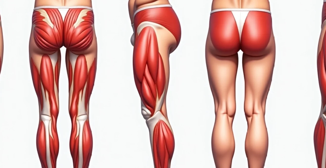
The quadriceps femoris represents the largest muscle group in your thigh, comprising four distinct heads that work collectively to extend the knee and flex the hip. When examining muscle depressions, understanding the normal anatomical boundaries becomes crucial for distinguishing pathological changes from natural variations in muscle architecture. The rectus femoris, vastus lateralis, vastus medialis, and vastus intermedius each contribute to the overall contour of your anterior thigh, and disruptions to any of these components can result in visible depressions or asymmetries.
Muscle depressions typically present as localised areas where the normal convex surface of the thigh becomes concave or flattened. These changes may develop gradually over weeks or months, particularly in cases involving muscle atrophy or denervation, or they may appear acutely following traumatic incidents. The location, size, and characteristics of these depressions provide valuable diagnostic clues about the underlying pathophysiology and potential treatment requirements.
Rectus femoris atrophy and visible muscle wasting characteristics
The rectus femoris, positioned centrally within the quadriceps group, frequently demonstrates visible wasting patterns that create characteristic depressions along the anterior thigh. This muscle’s superficial location makes atrophic changes readily apparent, particularly in individuals with lower body fat percentages. Rectus femoris atrophy often results from disuse, neurological impairment, or age-related sarcopenia, leading to a flattened or concave appearance where the muscle belly should maintain its normal bulk.
Vastus lateralis indentation: Location-Specific assessment markers
The vastus lateralis, extending along the outer aspect of your thigh, commonly exhibits depression patterns following direct trauma or compartment syndrome complications. These indentations typically appear as longitudinal grooves or localised dimpling that disrupts the normal curved outline of the lateral thigh. Assessment of vastus lateralis depressions requires careful palpation to distinguish between superficial fascia changes and deeper muscle architecture alterations.
Sartorius muscle groove differentiation from pathological depressions
The sartorius muscle creates a natural groove that runs diagonally across your anterior thigh, which may be mistaken for pathological depression in some individuals. This normal anatomical landmark becomes more prominent with muscle development or fat reduction, creating a visible indentation that should not cause concern. Distinguishing between this physiological groove and pathological muscle wasting requires understanding the expected course and characteristics of the sartorius muscle boundary.
Fascial compartment syndrome resulting in anterior thigh deformities
Chronic compartment syndrome affecting the anterior thigh can lead to permanent muscle depressions through increased intracompartmental pressure and subsequent muscle necrosis. These depressions often appear as irregular, firm indentations that may be accompanied by altered sensation or functional impairment. The fascial boundaries of the anterior compartment can become fibrotic following severe compartment syndrome episodes, creating permanent architectural changes that manifest as visible deformities.
Traumatic aetiologies: Impact-Related muscle tissue damage and contusion sequelae
Traumatic injuries to the thigh region represent one of the most common causes of muscle depressions, particularly following direct impact injuries sustained during contact sports, motor vehicle accidents, or falls. The quadriceps muscle group’s exposed anterior position makes it particularly vulnerable to blunt force trauma, which can result in various degrees of muscle damage ranging from minor contusions to severe crushing injuries. Understanding the progression of traumatic muscle injuries helps predict the likelihood of permanent depressions and guides appropriate treatment strategies.
Muscle contusions affecting the thigh can range from mild bruising with minimal long-term effects to severe injuries that permanently alter muscle architecture and function.
The severity of traumatic muscle depressions depends on multiple factors, including the magnitude of the applied force, the duration of impact, and the individual’s muscle mass and conditioning at the time of injury. Athletes participating in contact sports such as rugby, football, or martial arts frequently experience thigh contusions that may heal with residual depressions. The healing process involves complex inflammatory cascades and tissue remodelling that can sometimes result in irregular scar formation and permanent changes to muscle contour.
Haematoma resolution phases and residual tissue depression formation
Large haematomas within the quadriceps muscle undergo predictable resolution phases that may culminate in residual depressions or irregularities. The initial bleeding phase creates localised swelling and distension, followed by organisation and reabsorption of the clotted blood. During the remodelling phase, fibrous tissue may replace normal muscle architecture, creating areas of decreased volume that manifest as visible depressions. This process typically occurs over several weeks to months following the initial injury.
Myofibre rupture patterns in contact sports injuries
Direct impact injuries commonly cause myofibre rupture patterns that heal with varying degrees of scarring and architectural disruption. Grade II and III muscle strains involving significant fibre disruption may heal with irregular collagen deposition that creates localised depressions or thickened areas. The healing response involves satellite cell activation and new myofibre formation, but the regenerated muscle may not perfectly replicate the original architecture, particularly following severe injuries.
Fat necrosis following blunt force trauma to quadriceps region
Blunt force trauma to the thigh can cause localised fat necrosis within the subcutaneous and intramuscular fat compartments, leading to permanent depressions as the necrotic tissue is reabsorbed. This process, known as traumatic fat necrosis, typically occurs several weeks following the initial injury and may create irregular contour changes that persist indefinitely. The affected areas often feel firm to palpation and may be associated with skin changes or discolouration.
Crush injury complications leading to permanent muscle architecture changes
Severe crush injuries to the thigh region can cause extensive muscle necrosis and subsequent architectural remodelling that results in significant depressions or deformities. These injuries often involve multiple muscle groups and may be complicated by compartment syndrome, rhabdomyolysis, or infection. The healing process frequently involves extensive fibrosis and muscle replacement with non-contractile tissue, creating permanent changes to thigh contour and function.
Neuromuscular pathology: denervation atrophy and motor unit dysfunction
Neurological conditions affecting the motor innervation of the quadriceps muscles can result in progressive muscle atrophy and visible depressions as functional muscle mass decreases over time. The femoral nerve provides primary innervation to the quadriceps group, and any disruption to this neural pathway can lead to denervation atrophy characterised by muscle wasting and weakness. Understanding the relationship between neural function and muscle maintenance helps explain why some thigh depressions develop gradually without obvious traumatic causes.
Denervation atrophy typically presents as progressive muscle wasting that begins subtly and becomes more pronounced over weeks to months. The affected muscles lose their normal bulk and tone, creating visible depressions that may be accompanied by weakness, fasciculations, or altered reflexes. Common causes include lumbar radiculopathy, femoral neuropathy, diabetic neuropathy, or more generalised conditions such as motor neuron disease or muscular dystrophy.
The pattern of muscle atrophy provides diagnostic clues about the level and nature of the neurological lesion. Proximal muscle weakness affecting the hip flexors and knee extensors may suggest lumbar plexus involvement, while more distal patterns might indicate peripheral nerve pathology. Electromyography and nerve conduction studies become essential diagnostic tools for characterising the underlying neuromuscular disorder and guiding appropriate treatment strategies.
Metabolic myopathies, including inflammatory conditions such as polymyositis or dermatomyositis, can cause muscle wasting patterns that create visible depressions in the thigh region. These conditions typically affect multiple muscle groups and may be associated with systemic symptoms such as fatigue, muscle pain, or skin changes. The muscle wasting in metabolic myopathies often shows a characteristic distribution pattern that helps distinguish these conditions from neurological causes of atrophy.
Diagnostic imaging protocols: MRI and ultrasound assessment techniques
Advanced imaging modalities play a crucial role in evaluating thigh muscle depressions, providing detailed visualisation of muscle architecture, identifying underlying pathology, and guiding treatment decisions. Magnetic resonance imaging (MRI) represents the gold standard for assessing muscle injuries, offering superior soft tissue contrast and the ability to evaluate both acute and chronic changes within the quadriceps complex. Understanding the strengths and limitations of different imaging approaches helps clinicians select the most appropriate diagnostic strategy for individual patients.
MRI sequences specifically designed for muscle evaluation include T1-weighted images for anatomical detail, T2-weighted images for identifying oedema or inflammation, and STIR (Short Tau Inversion Recovery) sequences for suppressing fat signal and highlighting pathological changes. These imaging protocols can distinguish between acute muscle injuries, chronic scarring, fat infiltration, and other pathological processes that may contribute to visible depressions. Multi-planar imaging allows comprehensive assessment of muscle boundaries and relationships to surrounding structures.
Modern MRI techniques can detect subtle changes in muscle architecture that may not be apparent on clinical examination, providing valuable insights into the underlying pathophysiology of thigh depressions.
Ultrasound imaging offers several advantages for evaluating thigh muscle depressions, including real-time assessment, dynamic evaluation during muscle contraction, and cost-effectiveness compared to MRI. High-frequency ultrasound probes provide excellent resolution for superficial muscle evaluation, allowing assessment of muscle fibre integrity, scar tissue formation, and the presence of fluid collections or haematomas. The ability to perform comparative imaging of both thighs helps identify subtle asymmetries that may not be apparent clinically.
Specialised imaging techniques such as diffusion tensor imaging (DTI) or magnetic resonance neurography may be employed when neurological causes are suspected. These advanced modalities can assess nerve integrity and identify sites of compression or injury that may contribute to muscle denervation and subsequent atrophy. The integration of clinical findings with appropriate imaging results provides the foundation for accurate diagnosis and treatment planning.
Conservative treatment modalities: physiotherapy interventions and muscle rehabilitation
Conservative management approaches for thigh muscle depressions focus on addressing underlying causes, promoting muscle recovery, and preventing further deterioration through targeted therapeutic interventions. The specific treatment strategy depends on the underlying aetiology, with traumatic injuries requiring different approaches compared to neurological or metabolic causes. Physiotherapy interventions form the cornerstone of conservative management, emphasising progressive loading, functional restoration, and patient education regarding long-term muscle health maintenance.
Progressive resistance training represents a fundamental component of muscle rehabilitation, designed to stimulate hypertrophy and improve functional strength in affected muscles. The training programme must be carefully calibrated to the individual’s current capacity and underlying pathology, with gradual increases in load and complexity as healing progresses. Eccentric strengthening exercises may be particularly beneficial for promoting muscle remodelling and addressing architectural abnormalities that contribute to visible depressions.
Manual therapy techniques, including soft tissue mobilisation and myofascial release, can address restrictions in fascial planes that may contribute to muscle dysfunction and altered contours. These interventions help restore normal tissue extensibility and promote optimal muscle mechanics during functional activities. Therapeutic modalities such as ultrasound therapy, electrical stimulation, or photobiomodulation may enhance healing responses and promote tissue remodelling in appropriate cases.
- Progressive loading protocols tailored to individual recovery phases
- Functional movement retraining to address compensatory patterns
- Education regarding activity modification and injury prevention
- Ongoing monitoring and adjustment of treatment parameters
Nutritional optimisation plays a crucial supporting role in muscle recovery, with adequate protein intake being essential for muscle protein synthesis and tissue repair. Specific nutrients such as creatine, leucine, and vitamin D may provide additional benefits for muscle recovery and adaptation. Addressing any underlying metabolic or nutritional deficiencies becomes particularly important when treating muscle atrophy or delayed healing responses.
Medical emergency indicators: compartment syndrome recognition and immediate referral criteria
Recognising the signs and symptoms of acute compartment syndrome represents a critical clinical skill, as this condition constitutes a surgical emergency requiring immediate intervention to prevent permanent muscle necrosis and functional loss. Acute compartment syndrome occurs when increased pressure within the muscle compartments compromises blood flow and tissue perfusion, leading to rapid muscle death if left untreated. Understanding the classic presentation patterns and emergency referral criteria can be life-changing for affected individuals.
The cardinal symptoms of acute compartment syndrome include severe pain that appears disproportionate to the apparent injury, pain that worsens with passive stretching of the affected muscles, and progressive neurological symptoms such as numbness or weakness. The affected thigh typically becomes tense and swollen, with decreased sensation in areas supplied by nerves traversing the affected compartment. Pulselessness and paralysis represent late signs that indicate irreversible tissue damage and poor prognosis for functional recovery.
Time represents the most critical factor in compartment syndrome management, with delays in surgical decompression leading to irreversible muscle necrosis and permanent functional impairment.
Compartment pressure measurement using specialised monitoring equipment provides objective confirmation of the diagnosis when clinical signs are equivocal. Normal compartment pressures typically remain below 15 mmHg, with pressures exceeding 30 mmHg or within 30 mmHg of the diastolic blood pressure indicating the need for emergency fasciotomy. However, clinical judgment should supersede pressure measurements when classic symptoms are present, as normal pressure readings do not exclude the diagnosis in all cases.
Chronic compartment syndrome presents a more subtle clinical picture, with symptoms typically occurring during or after exercise and resolving with rest. This condition may contribute to gradual muscle changes and depressions through repeated episodes of elevated compartment pressure and subsequent tissue ischaemia. Recognition of chronic compartment syndrome requires a high index of suspicion and may necessitate dynamic pressure testing during provocative exercise to confirm the diagnosis.
Emergency referral becomes necessary when patients present with acute onset severe thigh pain, particularly following trauma or intensive exercise. Associated symptoms such as altered sensation, weakness, or signs of systemic illness warrant immediate medical evaluation. Healthcare providers should maintain a low threshold for emergency referral when compartment syndrome is suspected, as the consequences of delayed treatment far outweigh the risks of unnecessary surgical evaluation. Any patient presenting with progressive muscle weakness, altered sensation, or functional impairment in association with thigh depressions should receive prompt specialist assessment to exclude underlying neurological or vascular pathology requiring urgent intervention.