Discovering a hard lump beneath your caesarean section incision during the early postpartum period can understandably cause significant anxiety for new mothers. At two weeks post-surgery, your body remains in the active healing phase, and various physiological processes can manifest as palpable masses or areas of firmness along the surgical site. Understanding the normal wound healing trajectory and potential complications helps distinguish between expected recovery phenomena and situations requiring immediate medical attention.
The formation of lumps, bumps, or areas of induration near a fresh C-section incision represents a relatively common occurrence that affects approximately 15-20% of women during the initial healing phase. These manifestations can arise from multiple underlying causes, ranging from normal inflammatory responses to more serious complications such as haematoma formation or early infection development. Recognising the difference between normal healing processes and pathological conditions becomes crucial for appropriate management and optimal recovery outcomes.
Caesarean section wound healing pathophysiology and normal recovery timeline
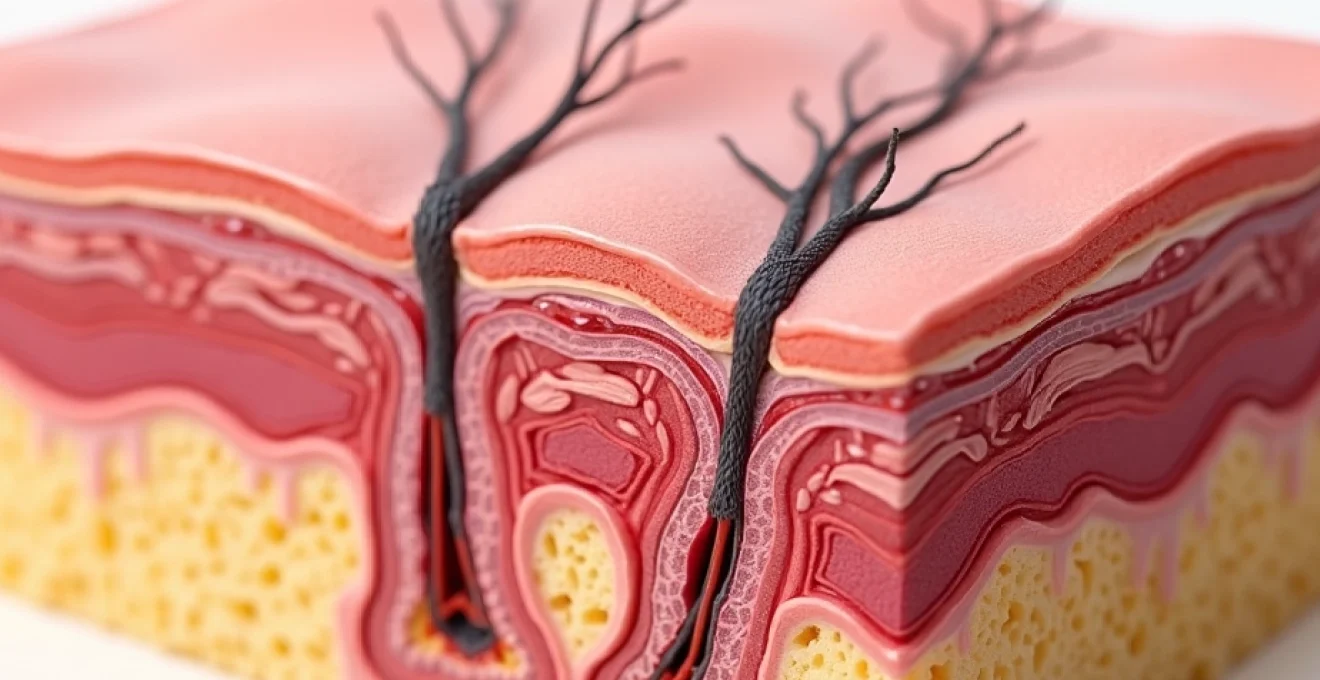
The caesarean section procedure involves creating incisions through multiple anatomical layers, including skin, subcutaneous tissue, rectus fascia, peritoneum, and uterine wall. Each of these tissue planes follows distinct healing patterns and timelines, contributing to the complex recovery process that extends well beyond the visible surface wound. During the initial two-week period, your body orchestrates an intricate cascade of biological events designed to restore tissue integrity and prevent infection.
Primary intention healing process in pfannenstiel incision sites
The horizontal Pfannenstiel incision, commonly known as the “bikini cut,” heals through primary intention when surgical edges are properly approximated with sutures or staples. This healing method involves direct tissue-to-tissue contact, minimising scar formation and reducing recovery time compared to secondary healing processes. The initial 48-72 hours witness rapid cellular migration and early fibrin clot formation, creating the foundation for subsequent tissue repair.
Primary intention healing relies heavily on adequate blood supply and minimal tissue tension to achieve optimal results. During the first two weeks, you may notice varying degrees of swelling, redness, and firmness as your body mobilises inflammatory mediators and growth factors essential for tissue regeneration. These changes often create palpable areas that can feel concerning but represent normal physiological responses.
Inflammatory phase characteristics during first 72 hours Post-Surgery
The inflammatory phase represents the body’s immediate response to surgical trauma, characterised by vasodilation, increased vascular permeability, and white blood cell recruitment to the wound site. During this critical period, you may experience localised warmth, swelling, and discomfort as inflammatory mediators such as histamine and prostaglandins coordinate the healing response. These symptoms typically peak within the first 48-72 hours before gradually subsiding.
Inflammatory exudate accumulation can create areas of firmness or fullness beneath the incision line, particularly in patients with robust immune responses or those experiencing mild complications. The presence of surgical drains can influence inflammatory patterns, though most caesarean sections do not require drainage systems unless specific risk factors are present.
Proliferative phase tissue regeneration at 7-14 days
Between days seven and fourteen post-surgery, the proliferative phase dominates the healing process as fibroblasts begin synthesising new collagen fibres and blood vessels establish revascularisation pathways. This period often coincides with suture removal or dissolution, potentially creating temporary areas of weakness or irregularity along the incision line. New tissue formation can feel firm, nodular, or rope-like as collagen deposits accumulate in organised patterns.
The proliferative phase also witnesses significant changes in wound appearance and texture, with many women noticing increased firmness extending several centimetres beyond the visible incision. This expansion of palpable tissue represents normal healing processes rather than pathological conditions, though distinguishing between normal and abnormal findings requires careful assessment.
Collagen deposition and scar maturation patterns
Collagen synthesis and remodelling continue for months following surgery, creating dynamic changes in scar tissue composition and characteristics. During the initial two-week period, immature collagen predominates, contributing to the firm, sometimes lumpy texture many women experience. Type III collagen initially fills the wound space before gradually being replaced by stronger Type I collagen over subsequent months.
The maturation process involves continuous collagen turnover, with breakdown and synthesis occurring simultaneously to optimise scar strength and flexibility. Understanding this ongoing remodelling helps explain why scar characteristics change significantly over time, with initial firmness often softening and smoothing during the following year.
Post-operative haematoma formation and seroma development
Haematoma and seroma formation represent two of the most common causes of palpable lumps following caesarean delivery. These fluid collections develop when normal haemostasis fails or when lymphatic drainage becomes impaired, creating localised accumulations that manifest as firm masses beneath the surgical site. Understanding the pathophysiology and risk factors associated with these complications enables better recognition and management strategies.
Subcutaneous haematoma pathogenesis and risk factors
Subcutaneous haematomas develop when bleeding occurs in the space between the skin and underlying fascial layers, often resulting from inadequate haemostasis during wound closure or post-operative vessel disruption. These collections typically present as firm, sometimes tender masses that may exhibit characteristic bluish discolouration resembling large bruises. The size and extent of subcutaneous haematomas can vary significantly , from small, localised collections to extensive masses spanning the entire incision length.
Several factors increase haematoma risk, including anticoagulant medications, bleeding disorders, hypertension, and advanced maternal age. Patients with previous abdominal surgeries or adhesions may experience increased bleeding due to tissue manipulation difficulties during the procedure. Recognising these risk factors helps healthcare providers implement appropriate monitoring and intervention strategies.
Seroma accumulation in camper’s fascia layer
Seromas form when lymphatic fluid accumulates in potential spaces created during surgical dissection, most commonly in Camper’s fascia layer of the subcutaneous tissue. Unlike haematomas, seromas contain clear or straw-coloured fluid and typically develop gradually over several days to weeks following surgery. The palpable characteristics often feel softer and more fluctuant compared to the firm consistency of haematomas.
Seroma development correlates strongly with the extent of subcutaneous dissection required during surgery, with complicated deliveries or repeat caesarean sections carrying higher risks. Drainage techniques and closure methods can influence seroma formation rates , though most small collections resolve spontaneously without intervention.
Rectus sheath haematoma following muscle retraction
Rectus sheath haematomas represent deeper bleeding complications occurring within or adjacent to the rectus abdominis muscle and its surrounding fascia. These collections often present as firm, fixed masses that may be tender to palpation and can cause significant discomfort during movement or coughing. The deeper location makes these haematomas more challenging to detect through routine examination alone.
Muscle retraction techniques used during caesarean delivery can occasionally result in small vessel injury or muscle fibre tears, predisposing to haematoma formation. The confined space within the rectus sheath can lead to increased pressure and pain as the collection expands, sometimes requiring more aggressive management approaches.
Vascular compromise and inferior epigastric artery injury
The inferior epigastric artery system supplies significant portions of the anterior abdominal wall and can be inadvertently injured during caesarean section procedures, particularly when anatomical variants exist or when surgical exposure proves challenging. Injury to these vessels can result in significant bleeding and haematoma formation, often requiring surgical intervention for resolution.
Vascular compromise may also affect wound healing through reduced tissue perfusion, potentially contributing to delayed healing, increased infection risk, or abnormal scar formation. Early recognition of vascular complications becomes crucial for preventing more serious sequelae and ensuring optimal recovery outcomes.
Incisional hernia development through fascial dehiscence
Incisional hernia formation represents a serious long-term complication that can occasionally present as early as two weeks post-surgery, though most cases develop gradually over months to years. The condition occurs when fascial layers fail to heal properly, creating weakened areas through which abdominal contents can protrude. Early hernia development typically results from technical factors during closure, patient-related risk factors, or post-operative complications that compromise healing.
The incidence of incisional hernia following caesarean section ranges from 0.2% to 2.0%, with higher rates observed in patients with multiple risk factors such as obesity, diabetes, smoking, or wound complications. Early detection of fascial dehiscence allows for prompt intervention and potentially better long-term outcomes compared to delayed recognition and treatment.
Distinguishing between normal post-operative swelling and early hernia formation requires careful examination techniques and sometimes imaging studies. The presence of a reducible mass that becomes more prominent with coughing or straining suggests possible hernia development, warranting immediate medical evaluation. Risk factors for early hernia formation include inadequate fascial closure, suture material failure, wound infection, and excessive post-operative strain on the surgical site.
Understanding the biomechanical factors contributing to fascial failure helps guide both prevention strategies and early intervention approaches for optimal patient outcomes.
Surgical site infection manifestations and abscess formation
Surgical site infections (SSI) affect approximately 3-5% of women following caesarean delivery and can present with various manifestations ranging from superficial cellulitis to deep abscesses requiring surgical drainage. The development of palpable lumps or masses at two weeks post-surgery may indicate evolving infectious processes that require prompt recognition and treatment to prevent serious complications.
Superficial incisional SSI clinical presentation
Superficial incisional infections involve only the skin and subcutaneous tissue layers, typically presenting with characteristic signs of inflammation including erythema, warmth, tenderness, and sometimes purulent drainage. These infections may create areas of induration or firm swelling that can be mistaken for normal healing processes during the early post-operative period. The key distinguishing features include progressive worsening of symptoms rather than gradual improvement expected during normal recovery.
Temperature elevation, though not universal, often accompanies superficial SSI development and may be the first systemic sign of infection. Local pain that increases rather than decreases over time, particularly when associated with spreading erythema, should raise suspicion for infectious complications requiring antibiotic therapy.
Deep incisional infection involving rectus fascia
Deep incisional infections extend beyond the subcutaneous layers to involve fascial planes and potentially the rectus muscle itself, creating more serious clinical scenarios requiring aggressive management approaches. These infections often present with systemic symptoms including fever, malaise, and elevated white blood cell counts, in addition to local signs of inflammation and possible wound drainage.
The presence of deep infection can compromise fascial integrity and increase the risk of subsequent hernia formation or wound dehiscence. Early identification and treatment of deep SSI becomes crucial for preventing these serious long-term complications and ensuring optimal surgical outcomes.
Staphylococcus aureus and streptococcus pyogenes pathogenicity
Staphylococcus aureus represents the most common pathogen causing post-caesarean wound infections, with methicillin-resistant strains (MRSA) posing particular treatment challenges in some patient populations. The organism’s virulence factors enable tissue invasion and abscess formation, often creating characteristic palpable masses with central fluctuance and surrounding induration.
Streptococcus pyogenes, though less common, can cause rapidly spreading cellulitis with minimal pus formation but significant systemic toxicity. The clinical presentation may include rapidly expanding erythema, severe pain, and systemic symptoms that can progress to life-threatening complications if not recognised and treated promptly.
Polymicrobial infection patterns in Post-Caesarean wounds
Many post-caesarean wound infections involve multiple bacterial species, particularly when bowel flora contamination occurs during surgery or when predisposing factors such as diabetes or immunosuppression exist. Polymicrobial infections often present with mixed clinical features and may require broader antibiotic coverage compared to single-pathogen infections.
The complexity of polymicrobial infections can make diagnosis and treatment more challenging, often requiring culture-guided antibiotic selection and sometimes surgical debridement for optimal outcomes. Understanding the polymicrobial nature of many post-surgical infections helps guide empirical treatment decisions while awaiting culture results.
Diagnostic assessment using ultrasound and physical examination
Accurate diagnosis of lumps or masses developing after caesarean section requires systematic evaluation combining detailed physical examination with appropriate imaging studies when indicated. Ultrasound imaging has emerged as the preferred initial diagnostic modality due to its non-invasive nature, excellent soft tissue resolution, and ability to distinguish between solid masses and fluid collections. The technique proves particularly valuable for evaluating suspected haematomas, seromas, or abscesses while avoiding radiation exposure concerns in breastfeeding mothers.
Physical examination techniques for post-caesarean lumps should include assessment of size, consistency, mobility, tenderness, and relationship to surrounding structures. Palpation should be performed gently but systematically, noting whether masses are superficial or deep, fixed or mobile, and whether they demonstrate fluctuance suggesting fluid content. The timing of symptom development and associated clinical features provide crucial diagnostic clues that help differentiate between various potential causes.
Color Doppler ultrasound can provide additional information about vascular patterns within suspicious masses, helping distinguish between inflammatory processes, organised haematomas, and potential malignancies. The technique also allows for real-time assessment of compressibility and internal echogenicity patterns that correlate with specific pathological processes.
Systematic diagnostic approaches combining clinical assessment with targeted imaging studies provide the foundation for accurate diagnosis and appropriate treatment planning in post-caesarean complications.
| Clinical Finding | Likely Diagnosis | Additional Features |
|---|---|---|
| Soft, fluctuant mass | Seroma | Non-tender, develops gradually |
| Firm, tender mass with bruising | Haematoma | Early post-operative onset |
| Hard, fixed mass with erythema | Abscess/Infection | Fever, systemic symptoms |
| Reducible mass, worse with strain | Incisional hernia | May be asymptomatic initially |
Clinical management protocols and intervention strategies
Management approaches for hard lumps developing beneath caesarean incisions depend heavily on accurate diagnosis and assessment of associated risk factors or complications. Conservative management remains appropriate for many conditions, particularly small haematomas or seromas that show signs of gradual resolution. However, certain presentations require immediate intervention to prevent serious complications or ensure optimal healing outcomes.
For suspected haematomas, initial management typically involves observation with serial examinations to monitor size and symptoms. Small, stable collections often resolve spontaneously over several weeks as the body reabsorbs the accumulated blood products. Larger haematomas or those associated with significant pain or expansion may require drainage procedures, either through needle aspiration or surgical evacuation depending on the collection’s characteristics and location.
Seroma management follows similar conservative principles initially, with many small collections resolving without intervention. However, large or persistent seromas may require aspiration to prevent complications such as infection or wound healing delays. Repeated aspirations are sometimes necessary, and some clinicians advocate for drain placement in cases of recurrent accumulation.
Infectious complications require prompt antibiotic therapy based on clinical presentation and, when available, culture results. Superficial infections often respond well to oral antibiotics with close monitoring, while deeper infections may require intravenous therapy and surgical drainage. The choice of antibiotic coverage should consider local resistance patterns and patient-specific risk factors.
Patient education plays a crucial role in management success, with clear instructions about warning signs requiring immediate medical attention, wound care techniques, and activity restrictions during the healing period. <em
</em
Regular follow-up appointments allow healthcare providers to monitor healing progress and identify complications early when intervention proves most effective. Understanding that recovery timelines vary significantly between individuals helps set realistic expectations and reduces anxiety during the healing process.
Preventive strategies focus on optimising healing conditions through proper nutrition, adequate rest, gentle mobilisation as tolerated, and adherence to post-operative care instructions. Maintaining stable blood sugar levels in diabetic patients, ensuring adequate protein intake for tissue repair, and avoiding activities that place excessive strain on the healing tissues all contribute to optimal recovery outcomes.
When surgical intervention becomes necessary, the specific approach depends on the underlying pathology and patient factors. Haematoma evacuation may be performed under local anaesthesia for superficial collections, while deeper abscesses often require formal surgical drainage with general anaesthesia. Incisional hernia repair typically involves mesh reinforcement to reduce recurrence rates, though the timing of such procedures must balance healing considerations with symptom severity.
The key to successful management lies in early recognition, appropriate diagnosis, and tailored treatment approaches that consider individual patient factors and specific pathological processes.
Multidisciplinary care involving obstetricians, general surgeons, infectious disease specialists, and wound care nurses may be necessary for complex cases or those involving multiple complications. This collaborative approach ensures comprehensive assessment and optimal treatment coordination while maintaining focus on the patient’s overall well-being and recovery goals.
Long-term monitoring remains important even after initial resolution of acute complications, as some conditions such as incisional hernias may develop or recur months to years after the initial surgery. Patient education about long-term warning signs and the importance of maintaining healthy lifestyle factors supports sustained recovery and early detection of delayed complications.
Documentation of complications and treatment responses contributes to improved understanding of risk factors and optimal management strategies for future cases. This systematic approach to learning from clinical experience helps refine treatment protocols and improve outcomes for all patients undergoing caesarean delivery.
Remember that while discovering a hard lump beneath your caesarean incision can feel alarming, most cases have treatable causes with excellent prognosis when properly managed. Working closely with your healthcare team, following recommended treatment plans, and maintaining open communication about symptoms and concerns provides the best foundation for successful recovery and optimal long-term outcomes.