Discovering a lump at the back of your neck near the hairline can be concerning, particularly when it appears suddenly or grows over time. The posterior cervical region, where the neck meets the occipital area of the skull, is susceptible to various conditions that can manifest as palpable masses. Understanding the potential causes of these lumps is crucial for determining appropriate medical evaluation and treatment strategies.
The anatomical complexity of this region, with its rich network of lymph nodes, sebaceous glands, hair follicles, and subcutaneous tissues, creates multiple pathways for lump formation. Most lumps in this area are benign, arising from normal physiological processes or minor inflammatory responses. However, the location’s proximity to critical structures and lymphatic drainage pathways means that some masses may require immediate medical attention.
Healthcare professionals encounter posterior neck lumps frequently in clinical practice, with studies indicating that approximately 80% of neck masses in adults prove to be benign. The key to proper management lies in understanding the distinguishing characteristics of various conditions that can present as lumps in this anatomically significant region.
Benign subcutaneous masses at the posterior cervical region
Benign subcutaneous masses represent the most common cause of lumps appearing at the back of the neck near the hairline. These non-cancerous growths develop within the layers of tissue beneath the skin and typically present with characteristic features that help distinguish them from more serious conditions. The benign nature of these masses doesn’t diminish their clinical significance, as they can cause discomfort, cosmetic concerns, or secondary complications if left untreated.
Sebaceous cyst formation and keratin accumulation
Sebaceous cysts, also known as epidermoid cysts, frequently develop in the posterior neck region due to the high concentration of sebaceous glands and hair follicles in this area. These cysts form when sebaceous ducts become blocked, leading to the accumulation of keratin and sebaceous material within a capsule-like structure. The result is a smooth, round mass that feels firm to palpation and may exhibit slight mobility under the skin.
The characteristic features of sebaceous cysts include their slow growth pattern, typically developing over months or years, and their tendency to maintain a consistent size once formed. These cysts often present with a central punctum , a small dark spot representing the blocked follicular opening, which serves as a diagnostic hallmark. Patients frequently report that these masses become more prominent during periods of hormonal fluctuation or increased sebaceous gland activity.
Lipoma development in the suboccipital triangle
Lipomas represent another common benign cause of posterior neck lumps, particularly in the suboccipital triangle region where fatty tissue naturally accumulates. These soft tissue tumours consist of mature adipocytes encapsulated by a thin fibrous membrane, creating a characteristic soft, doughy texture that distinguishes them from other mass types. Lipomas typically exhibit excellent mobility under the skin and rarely cause pain unless they compress adjacent structures.
The suboccipital location is particularly prone to lipoma development due to the natural anatomical arrangement of fatty tissues in this region. Multiple lipomas can occur simultaneously , a condition known as familial multiple lipomatosis, which may have genetic predisposition factors. These masses generally grow slowly and may reach considerable sizes before patients seek medical attention, often measuring several centimetres in diameter.
Dermoid cyst pathophysiology at the occipital bone junction
Dermoid cysts represent developmental anomalies that can manifest as lumps at the posterior hairline, particularly near the occipital bone junction. These congenital lesions result from the entrapment of ectodermal elements during embryonic development, creating cysts lined with stratified squamous epithelium containing hair follicles, sebaceous glands, and sometimes teeth or cartilage. The location at the occipital region corresponds to embryonic closure lines where such entrapment commonly occurs.
Unlike acquired cysts, dermoid cysts may be present from birth but often remain unnoticed until later in life when growth or inflammation brings them to clinical attention. These masses typically feel firmer than sebaceous cysts and may demonstrate attachment to deeper structures, including the underlying periosteum or even intracranial extensions in rare cases.
Pilomatrixoma: calcifying epithelioma of malherbe
Pilomatrixoma, historically termed calcifying epithelioma of Malherbe, represents a benign tumour of hair matrix cells that can present as firm lumps in the posterior neck region. This condition predominantly affects children and young adults, with a characteristic presentation of a hard, sometimes calcified mass that may feel stone-like to palpation. The tumour derives from primitive cells of the hair matrix and typically demonstrates slow growth with potential for calcification over time.
The diagnostic challenge with pilomatrixoma lies in its firm consistency, which can mimic more serious conditions. However, the characteristic “tent sign” – where the overlying skin can be elevated while the mass remains fixed to deeper structures – helps differentiate this condition from other causes of posterior neck lumps.
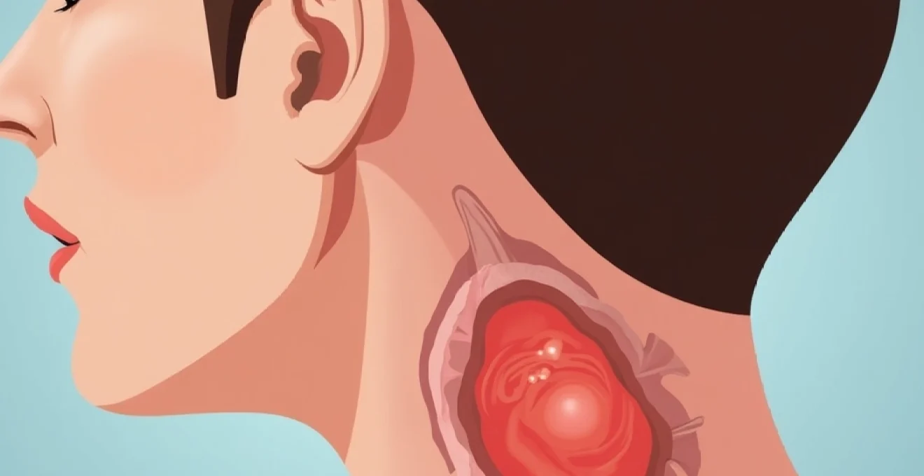
Lymphatic system abnormalities and cervical lymphadenopathy
The posterior cervical region contains numerous lymph nodes that form part of the body’s immune surveillance system. These lymph nodes can become enlarged due to various causes, ranging from benign reactive processes to malignant infiltration. Understanding the patterns of cervical lymphadenopathy is crucial for healthcare providers, as the characteristics of enlarged nodes often provide important diagnostic clues about underlying conditions.
Reactive lymph node enlargement in suboccipital chain
Reactive lymphadenopathy represents the most common cause of lymph node enlargement in the posterior cervical region, typically occurring in response to local or systemic infections. The suboccipital lymph nodes, located at the junction of the neck and skull, drain the posterior scalp region and frequently enlarge in response to scalp infections, seborrheic dermatitis, or upper respiratory tract infections. These nodes characteristically feel soft to rubbery and may be tender to palpation during acute inflammatory phases.
The temporal relationship between infection and node enlargement provides important diagnostic information. Reactive nodes typically appear within days of the precipitating infection and gradually decrease in size as the underlying condition resolves. However, complete resolution may take several weeks to months, particularly in cases of chronic or recurrent infections.
Hodgkin’s lymphoma manifestation in posterior cervical nodes
Hodgkin’s lymphoma can present with enlarged lymph nodes in the posterior cervical chain, though this pattern is less common than anterior cervical or mediastinal involvement. When posterior cervical nodes are affected, they typically present as firm, non-tender masses that demonstrate progressive enlargement over weeks to months. The characteristic feature of Hodgkin’s lymphoma is its tendency to spread in an orderly, contiguous manner from one node group to adjacent groups.
Patients with Hodgkin’s lymphoma affecting posterior cervical nodes may experience constitutional symptoms including fever, night sweats, and unintentional weight loss – collectively known as B symptoms. The presence of these systemic symptoms significantly influences staging and treatment decisions, making their recognition crucial for optimal patient management.
Non-hodgkin’s lymphoma: diffuse large B-Cell presentation
Diffuse large B-cell lymphoma, the most common subtype of non-Hodgkin’s lymphoma, can manifest as rapidly growing masses in the posterior cervical region. Unlike Hodgkin’s lymphoma, non-Hodgkin’s varieties tend to present in a more random, non-contiguous pattern and often involve multiple node groups simultaneously. These tumours typically feel firm to hard on palpation and may demonstrate rapid growth over days to weeks.
The aggressive nature of diffuse large B-cell lymphoma means that patients often present with advanced disease at diagnosis. Posterior cervical involvement may be accompanied by involvement of other node groups, extranodal sites, or both, requiring comprehensive staging evaluation to determine disease extent and appropriate treatment strategies.
Kikuchi-fujimoto disease: histiocytic necrotising lymphadenitis
Kikuchi-Fujimoto disease, also known as histiocytic necrotising lymphadenitis, represents a rare but important cause of posterior cervical lymphadenopathy. This self-limiting condition predominantly affects young adults, particularly women, and presents with painful lymph node enlargement that may be accompanied by fever and malaise. The disease process involves extensive necrosis within affected lymph nodes, leading to their characteristic firm, tender consistency.
The diagnosis of Kikuchi-Fujimoto disease often requires lymph node biopsy, as the clinical presentation can mimic lymphoma or other serious conditions. The histopathological findings are pathognomonic , showing characteristic necrotising inflammation without neutrophilic infiltration. The condition typically resolves spontaneously over several months without specific treatment.
Tuberculosis lymphadenitis and mycobacterial infections
Tuberculosis lymphadenitis, historically known as scrofula, can present as chronic lymph node enlargement in the posterior cervical region, particularly in immunocompromised patients or those from endemic areas. The condition results from haematogenous spread of Mycobacterium tuberculosis to lymph nodes, where the organisms establish chronic granulomatous inflammation. These nodes typically feel firm and may demonstrate matting or adherence to surrounding structures.
Atypical mycobacterial infections can also cause similar presentations, particularly in children and immunocompromised adults. The differentiation between typical and atypical mycobacterial infections requires specific diagnostic testing, including acid-fast bacilli staining, culture, and molecular identification techniques. Treatment approaches differ significantly between these conditions, making accurate diagnosis essential.
Inflammatory conditions and infectious aetiologies
Inflammatory and infectious processes represent significant causes of lump formation in the posterior neck region. These conditions often present with acute onset symptoms and may be accompanied by systemic signs of infection or inflammation. Understanding the pathophysiology and presentation patterns of these conditions is essential for timely diagnosis and appropriate treatment intervention.
Bacterial cellulitis and staphylococcus aureus infections
Bacterial cellulitis in the posterior neck region commonly results from Staphylococcus aureus or Streptococcus pyogenes infections, often secondary to minor trauma, hair follicle damage, or pre-existing skin conditions. The infection typically begins in the superficial tissues but can progress to involve deeper structures, creating palpable masses that feel warm, tender, and indurated. The characteristic erythematous border helps distinguish cellulitis from other causes of neck swelling.
Methicillin-resistant Staphylococcus aureus (MRSA) infections have become increasingly prevalent in both community and healthcare settings, requiring specific antibiotic selection based on culture and sensitivity results. The posterior neck region’s rich blood supply generally ensures good antibiotic penetration, but severe cases may require intravenous therapy or surgical intervention.
Hidradenitis suppurativa in the posterior neck region
Hidradenitis suppurativa can affect the posterior neck region, particularly in areas where apocrine glands are present and friction occurs. This chronic inflammatory condition involves the pilosebaceous unit and creates recurrent, painful nodules that may progress to form abscesses, sinus tracts, and scarring. The condition typically affects adults and demonstrates a predilection for areas subject to friction and moisture.
The diagnosis of hidradenitis suppurativa in the posterior neck requires recognition of the characteristic pattern of recurrent inflammation, often with a history of similar lesions in other typical locations such as the axillae or groin. Early intervention with appropriate medical therapy can prevent progression to severe scarring and functional impairment.
Folliculitis decalvans and chronic scalp inflammation
Folliculitis decalvans represents a chronic inflammatory condition affecting hair follicles in the posterior scalp region, potentially creating palpable inflammatory masses along the hairline. This neutrophilic scarring alopecia involves destruction of hair follicles with replacement by fibrous tissue, often resulting in permanent hair loss in affected areas. The condition can present as tender, inflammatory nodules that may be mistaken for other causes of posterior neck lumps.
The pathogenesis of folliculitis decalvans involves bacterial colonisation, typically with Staphylococcus aureus, combined with an abnormal host inflammatory response. Treatment requires long-term antibiotic therapy and anti-inflammatory measures to control the disease process and prevent further follicular destruction.
Carbuncle formation and furuncle clusters
Carbuncles represent clusters of interconnected furuncles that commonly develop in the posterior neck region due to the high density of hair follicles and frequent friction from clothing or sports equipment. These deep-seated bacterial infections typically involve multiple adjacent follicles, creating large, painful masses with multiple drainage points. The posterior neck location is particularly susceptible due to constant moisture from perspiration and mechanical irritation.
The development of carbuncles often indicates underlying predisposing factors such as diabetes mellitus, immunosuppression, or chronic skin conditions. Management requires systemic antibiotic therapy, often combined with surgical drainage for large or persistent lesions. Prevention strategies focus on maintaining proper hygiene and addressing underlying risk factors that predispose to recurrent infections.
Malignant neoplasms and metastatic disease processes
While malignant causes represent a minority of posterior neck lumps, their potential for serious consequences necessitates careful evaluation and prompt recognition. Malignant neoplasms in this region can arise as primary tumours or represent metastatic disease from distant sites. The characteristics that suggest malignancy include rapid growth, firm to hard consistency, fixation to underlying structures, and associated constitutional symptoms.
Primary malignancies of the posterior neck region include various sarcomas, particularly those arising from soft tissue or bone structures. Rhabdomyosarcoma, liposarcoma, and malignant fibrous histiocytoma can all present as rapidly growing masses in this location. These tumours typically demonstrate aggressive local growth patterns and may metastasise to regional lymph nodes or distant sites. Early recognition and referral to specialised oncology services is crucial for optimal patient outcomes.
Metastatic disease to the posterior cervical region can occur from various primary sites, including lung, breast, kidney, and gastrointestinal malignancies. The posterior cervical lymph nodes serve as potential sites for metastatic deposits, particularly from scalp or neck primary tumours. Squamous cell carcinoma of the scalp or posterior neck skin can metastasise to regional nodes, creating firm, non-tender masses that may be fixed to surrounding structures.
Melanoma represents a particularly concerning cause of posterior neck lumps, either as primary lesions arising from the scalp or neck skin, or as metastatic deposits in regional lymph nodes. The posterior neck and scalp are common sites for melanoma development, particularly in individuals with significant sun exposure or fair skin complexions. Any pigmented lesion demonstrating asymmetry, border irregularity, colour variation, diameter greater than 6mm, or evolution over time requires immediate dermatological evaluation.
The evaluation of potentially malignant posterior neck lumps requires systematic assessment including detailed history taking, physical examination, and appropriate imaging studies. Features that suggest malignancy include patient age over 40 years, smoking history, rapid growth over weeks rather than months, firm or hard consistency, fixation to deeper structures, and the presence of constitutional symptoms such as weight loss, fever, or night sweats. Prompt referral to appropriate specialists ensures timely diagnosis and treatment initiation.
Diagnostic imaging modalities and clinical assessment protocols
The evaluation of posterior neck lumps requires a systematic approach combining clinical assessment with appropriate diagnostic imaging modalities. The initial clinical evaluation focuses on determining the likelihood of malignancy and guiding subsequent investigation pathways. Physical examination characteristics, including size, consistency, mobility, and associated symptoms, provide crucial information for differential diagnosis and risk stratification.
Ultrasonography represents the initial imaging modality of choice for evaluating posterior neck lumps due to its accessibility, cost-effectiveness, and ability to differentiate between solid and cystic lesions. High-resolution ultrasound can distinguish between various tissue types, assess vascularity through Doppler studies, and guide fine-needle aspiration procedures when indicated. Characteristic ultrasound features help differentiate benign from malignant processes, with malignant nodes typically demonstrating loss of normal architecture, increased vascularity, and irregular borders.
Computed tomography (CT) scanning provides detailed cross-sectional imaging that proves particularly valuable for assessing the relationship between masses and surrounding anatomical structures. CT imaging excels at detecting calcification within masses, evaluating bony involvement, and identifying deep extension patterns that may not be apparent on physical examination. Contrast-enhanced CT studies can further delineate vascular relationships and enhance the visualisation of inflammatory or neoplastic processes.
Magnetic resonance imaging (MRI) offers superior soft tissue contrast resolution, making it the optimal imaging modality for evaluating complex masses or those with suspected intracranial extension. MRI sequences can differentiate between various tissue types, identify cystic components, and assess for perineural spread or other patterns of malignant extension. The multiplanar imaging capabilities of MRI provide comprehensive three-dimensional assessment of mass relationships to critical structures.
Fine-needle aspiration (FNA) cytology represents a minimally invasive diagnostic procedure that can provide definitive tissue diagnosis for many posterior neck masses. Under ultrasound guidance, FNA can safely sample both superficial and deep masses while avoiding critical neurovascular structures. The cytological findings combined with clinical correlation often provide sufficient information for definitive diagnosis and treatment planning, particularly for lymphomatous processes or metastatic disease.
Core needle biopsy may be indicated when FNA fails to provide diagnostic tissue or when architectural assessment is crucial for accurate diagnosis. This technique provides larger tissue samples that preserve histological architecture, enabling more comprehensive pathological evaluation including immunohistochemical studies and molecular analysis when indicated. The posterior neck location generally allows safe access for core biopsy procedures under imaging guidance.
Treatment algorithms and surgical intervention strategies
The management of posterior neck lumps requires individualised treatment approaches based on accurate diagnosis, patient factors, and lesion characteristics. Treatment algorithms begin with risk stratification based on clinical and imaging findings, followed by definitive diagnostic procedures when indicated. The therapeutic approach must balance the need for definitive treatment with consideration of cosmetic outcomes and functional preservation.
Conservative management strategies apply to clearly benign lesions that do not cause symptoms or cosmetic concerns. Sebaceous cysts, small lipomas, and reactive lymph nodes often require only observation with periodic reassessment to monitor for changes in size or character. Patient education regarding warning signs ensures appropriate medical consultation if lesion characteristics change over time.
Surgical excision represents the definitive treatment for most benign solid masses causing symptoms or cosmetic concerns. The surgical approach depends on lesion location, size, and relationship to surrounding structures. Simple excision with primary closure suffices for most superficial lesions, while deeper masses may require more complex reconstruction techniques to achieve optimal cosmetic and functional outcomes.
For cystic lesions, complete excision including the cyst wall prevents recurrence and provides tissue for histopathological examination. Incomplete removal or simple drainage of cystic contents typically results in recurrence, making complete surgical excision the preferred approach when treatment is indicated. The timing of surgical intervention should consider patient preferences, lesion growth patterns, and potential for malignant transformation in specific lesion types.
Infectious and inflammatory conditions require targeted antimicrobial or anti-inflammatory therapy as the primary treatment modality. Bacterial cellulitis responds to appropriate antibiotic selection based on likely pathogens and local resistance patterns. Severe infections may require surgical drainage combined with antibiotic therapy, particularly when abscess formation occurs or when conservative management fails to achieve resolution.
Malignant lesions necessitate urgent referral to appropriate oncological specialists for comprehensive staging and multidisciplinary treatment planning. The treatment approach for malignant posterior neck masses typically involves surgical resection with appropriate margins, often combined with adjuvant chemotherapy or radiation therapy based on tumour type and stage. Early recognition and prompt referral significantly influence patient outcomes in malignant conditions.
Lymphomatous processes require haematological evaluation and staging studies to determine disease extent and appropriate treatment protocols. The treatment of lymphomas typically involves chemotherapy regimens specific to histological subtype and disease stage, with radiation therapy reserved for specific indications. The posterior cervical location rarely influences treatment selection but may affect radiation field planning when indicated.
Post-treatment surveillance protocols vary based on the underlying condition and treatment modality employed. Benign lesions treated with complete surgical excision typically require no further follow-up unless concerning symptoms develop. Malignant conditions require structured surveillance programs including periodic physical examination, imaging studies, and laboratory monitoring as appropriate for the specific tumour type and treatment received.
What role does patient education play in the management of posterior neck lumps? Comprehensive patient education ensures understanding of diagnosis, treatment options, and expected outcomes while emphasising the importance of compliance with follow-up recommendations. Patients should understand warning signs that warrant immediate medical attention, including rapid growth, development of constitutional symptoms, or changes in lesion characteristics that might suggest malignant transformation.
The multidisciplinary approach to posterior neck mass management often involves collaboration between primary care physicians, dermatologists, surgical specialists, and oncologists depending on the underlying diagnosis. Effective communication between healthcare providers ensures coordinated care delivery and optimal patient outcomes while minimising unnecessary procedures or delays in definitive treatment.