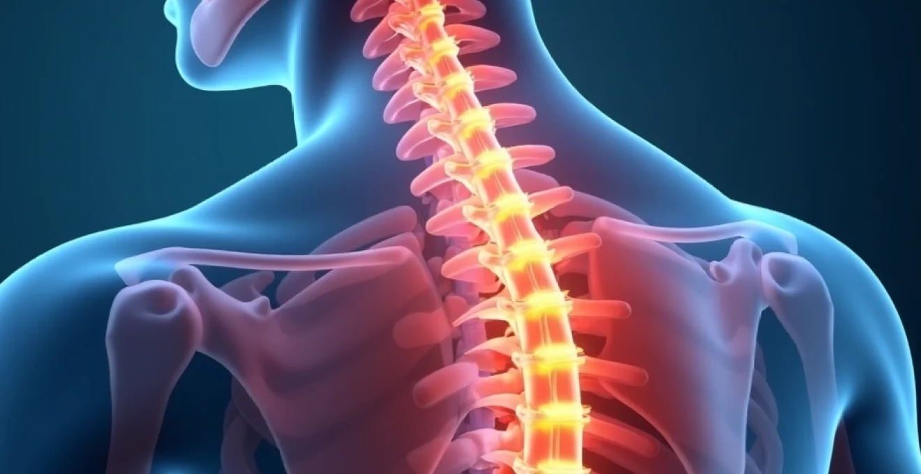
The thoracic spine’s mid-region, particularly the T5 and T6 vertebrae, represents a critical anatomical junction where stability meets mobility. These vertebrae, positioned between the shoulder blades, endure unique biomechanical stresses that can lead to various pain syndromes affecting millions of individuals worldwide. Understanding the specific causes of T5-T6 pain requires examining the complex interplay between structural anatomy, degenerative processes, and environmental factors that contribute to discomfort in this vital spinal region.
Pain originating from the T5 and T6 vertebrae often manifests as a deep, aching sensation that can radiate around the ribcage or present as sharp, stabbing discomfort during movement. The prevalence of thoracic spine pain, whilst lower than cervical or lumbar complaints, affects approximately 15-35% of the adult population at some point in their lives. The unique challenges presented by T5-T6 pathology stem from the region’s dual role in providing stability for the thoracic cage whilst accommodating the complex movements required for daily activities.
Anatomical structure and biomechanics of T5-T6 thoracic vertebrae
The T5 and T6 vertebrae occupy a pivotal position within the thoracic spine, serving as the structural foundation for the mid-thoracic region. These vertebrae demonstrate distinctive anatomical characteristics that differentiate them from their cervical and lumbar counterparts. Each vertebra features a robust vertebral body designed to bear significant axial loads, coupled with posteriorly oriented transverse processes that articulate directly with the corresponding ribs. The vertebral bodies of T5 and T6 are intermediate in size, larger than upper thoracic segments but smaller than lower thoracic levels, reflecting the graduated increase in load-bearing requirements throughout the spinal column.
The pedicles of T5 and T6 vertebrae are characteristically thick and strong, creating a protective corridor for the spinal cord whilst providing attachment points for the laminae and facet joints. The spinous processes project posteriorly and inferiorly, overlapping with adjacent vertebrae to create the distinctive thoracic kyphotic curve. This overlapping arrangement, known as the “shingle effect,” provides enhanced stability but can limit extension movements and contribute to specific pathological processes when disrupted.
Costovertebral joint articulation with fifth and sixth ribs
The costovertebral joints represent one of the most significant anatomical features distinguishing the T5 and T6 vertebrae from other spinal segments. The fifth and sixth ribs articulate with their corresponding vertebrae through two distinct joint surfaces: the costotransverse joint and the costovertebral joint proper. The costotransverse joint connects the rib tubercle to the transverse process, whilst the costovertebral joint links the rib head to the vertebral body and intervertebral disc.
These articulations are synovial joints surrounded by robust capsular ligaments that provide both stability and mobility. The biomechanical significance of these joints cannot be overstated, as they must accommodate the complex three-dimensional movements of respiration whilst maintaining structural integrity during trunk rotation and lateral flexion. When dysfunction occurs in these costovertebral articulations, it can manifest as localised pain that may be mistaken for muscular strain or referred cardiac symptoms.
Thoracic kyphosis curvature impact on T5-T6 alignment
The natural thoracic kyphotic curve places the T5 and T6 vertebrae near the apex of this posterior convexity, creating specific biomechanical challenges. This curvature, typically measuring between 20-40 degrees in healthy individuals, distributes compressive forces primarily to the anterior aspects of the vertebral bodies and intervertebral discs. The kyphotic alignment results in increased flexion moments across the T5-T6 segments, potentially predisposing these levels to anterior wedge compression and disc degeneration over time.
The kyphotic positioning also influences the orientation of the facet joints , which must accommodate both the curved spinal alignment and the rotational demands of thoracic movement. Abnormal variations in thoracic kyphosis, whether hyperkyphotic or hypokyphotic, can significantly alter the stress distribution patterns across the T5-T6 segments, potentially accelerating degenerative changes and contributing to pain syndromes.
Intervertebral disc composition and load distribution patterns
The T5-T6 intervertebral disc demonstrates unique compositional characteristics that reflect its position within the thoracic spine. The nucleus pulposus maintains a relatively high water content compared to lumbar discs, but lower than cervical levels, creating specific viscoelastic properties that influence load distribution. The annulus fibrosus consists of approximately 15-20 concentric lamellae, with collagen fibres oriented at alternating 30-degree angles to provide optimal resistance to rotational and shear forces.
The disc height at T5-T6 is proportionally smaller relative to the vertebral body size when compared to lumbar segments, creating a different mechanical environment. This reduced disc height contributes to the limited flexion-extension range of motion characteristic of the thoracic spine whilst maintaining adequate space for the spinal cord within the vertebral canal. The load distribution patterns across the T5-T6 disc vary significantly between static postures and dynamic activities, with compressive loads increasing substantially during forward flexion activities such as lifting or prolonged sitting.
Facet joint orientation and movement restrictions in Mid-Thoracic region
The facet joints at T5-T6 demonstrate a characteristic orientation that significantly influences movement patterns and potential pathology. These synovial joints are oriented approximately 60 degrees from the horizontal plane and 20 degrees from the frontal plane, creating an oblique articulation that permits rotation whilst restricting sagittal plane motion. This orientation is intermediate between the more horizontally oriented cervical facets and the more sagittally oriented lumbar facets.
The articular surfaces are covered with hyaline cartilage and enclosed within a synovial capsule that provides both lubrication and proprioceptive feedback. The facet joint capsules at T5-T6 are reinforced by the ligamentum flavum posteriorly and contain numerous mechanoreceptors that contribute to spinal proprioception and muscle activation patterns. When these joints become inflamed or degenerated, they can produce localised pain that may radiate along the intercostal nerve pathways, creating symptoms that extend around the thoracic cage.
Degenerative pathology and Age-Related changes in T5-T6 segments
Degenerative changes affecting the T5 and T6 vertebrae follow predictable patterns that correlate with both chronological aging and cumulative mechanical stress. The thoracic spine generally experiences less severe degenerative changes compared to the cervical and lumbar regions due to the stabilising influence of the rib cage. However, the T5-T6 segments can still develop significant pathology that contributes to pain and functional limitation. Research indicates that degenerative disc disease becomes increasingly prevalent after the fourth decade of life, with approximately 40% of individuals over 40 years demonstrating some degree of T5-T6 disc degeneration on advanced imaging studies.
The degenerative cascade typically begins with alterations in disc biochemistry, progressing through structural changes in the annulus fibrosus and nucleus pulposus before affecting adjacent vertebral structures. Understanding these progressive changes is crucial for clinicians as it helps explain the often insidious onset of symptoms and guides appropriate treatment strategies. The mid-thoracic location of T5-T6 creates a unique mechanical environment where the transition from the more mobile upper thoracic spine to the stiffer lower thoracic region can concentrate stresses and accelerate degenerative processes.
Thoracic disc degeneration and schmorl’s node formation
Disc degeneration at the T5-T6 level follows a characteristic pattern beginning with biochemical changes in the nucleus pulposus. The proteoglycan content decreases, leading to reduced water-binding capacity and altered viscoelastic properties. This process typically manifests as disc height loss, increased signal intensity on T2-weighted MRI sequences, and progressive annular fissuring. The reduced hydrostatic pressure within the nucleus allows for increased stress concentration on the annular fibres, potentially leading to tears and herniation.
Schmorl’s nodes represent a specific manifestation of disc degeneration where nuclear material herniates through the vertebral endplate into the adjacent vertebral body. These lesions are particularly common in the thoracic spine and can occur at T5-T6 due to the combination of kyphotic loading and endplate weakness. Whilst many Schmorl’s nodes remain asymptomatic, acute formation can cause significant pain and may be associated with vertebral body oedema visible on MRI. The presence of multiple Schmorl’s nodes may indicate underlying metabolic bone disease or genetic predisposition to endplate weakness.
Facet joint arthropathy and osteophyte development
Facet joint arthropathy at T5-T6 represents a common source of axial thoracic pain, particularly in individuals over 50 years of age. The degenerative process begins with cartilage fibrillation and progresses through subchondral sclerosis, joint space narrowing, and eventual osteophyte formation. The unique oblique orientation of thoracic facet joints means that degenerative changes can significantly impact rotational mobility whilst creating potential for nerve root irritation as osteophytes encroach upon the neural foramina.
The development of facet joint hypertrophy and osteophytosis can contribute to spinal canal stenosis, although this is less common in the thoracic spine compared to lumbar levels. However, the relatively smaller thoracic spinal canal means that even modest degrees of facet hypertrophy can create clinically significant stenosis. Synovial cysts arising from degenerated facet joints represent another potential complication, although these are rare in the thoracic spine compared to lumbar segments.
Ligamentum flavum hypertrophy and spinal canal stenosis
The ligamentum flavum undergoes characteristic age-related changes that can contribute to T5-T6 pathology. This elastic ligament normally maintains tension during spinal flexion whilst buckling during extension to prevent impingement on neural structures. With aging, the ligament loses elasticity due to increased collagen content and calcification, leading to thickening and reduced compliance. Hypertrophied ligamentum flavum can contribute to central canal stenosis and may compress the spinal cord during extension movements.
In the thoracic spine, ligamentum flavum hypertrophy is often associated with ossification of the posterior longitudinal ligament (OPLL), creating a “double crush” scenario for the spinal cord. This combination can produce myelopathic symptoms including gait disturbance, upper extremity weakness, and bladder dysfunction.
The relatively narrow thoracic spinal canal makes even modest degrees of ligamentum flavum hypertrophy potentially significant for spinal cord compression.
Vertebral body compression fractures and osteoporotic changes
Osteoporotic compression fractures commonly affect the T5 and T6 vertebrae, particularly in postmenopausal women and elderly individuals with metabolic bone disease. The thoracic spine’s natural kyphotic curvature creates a mechanical environment that predisposes to anterior wedge compression fractures under relatively minor loading conditions. These fractures can occur spontaneously or following minimal trauma such as coughing, sneezing, or lifting light objects in individuals with severe osteoporosis.
The clinical presentation of T5-T6 compression fractures varies from acute severe pain to chronic aching discomfort. Fresh fractures may be associated with vertebral body oedema and local tenderness, whilst chronic fractures can contribute to progressive kyphotic deformity and secondary muscular pain. Multiple compression fractures in the thoracic spine can significantly reduce pulmonary function by decreasing chest wall expansion and compromising respiratory mechanics.
Traumatic injuries and mechanical dysfunction of T5-T6 vertebrae
Traumatic injuries affecting the T5 and T6 vertebrae present unique challenges due to the region’s anatomical complexity and the potential for both spinal cord and thoracic organ involvement. The mid-thoracic spine’s relative stability, provided by the rib cage articulations, means that significant trauma is typically required to produce structural damage. However, when injuries do occur, they often involve multiple anatomical structures including the vertebrae, ribs, costovertebral joints, and surrounding musculature. Motor vehicle accidents account for approximately 45% of thoracic spine trauma, followed by falls from height (20%) and sports-related injuries (15%).
The mechanism of injury significantly influences the pattern of structural damage and subsequent pain presentation. Flexion-compression mechanisms typically produce anterior wedge compression fractures, whilst axial loading can result in burst fractures with potential neurological compromise. Rotation-flexion injuries may cause facet joint dislocations or fracture-dislocations, representing some of the most unstable thoracic spine injuries. Understanding these injury patterns is crucial for accurate diagnosis and appropriate management, as missed or delayed recognition can lead to progressive deformity and neurological deterioration.
Mechanical dysfunction at T5-T6 can also result from repetitive microtrauma rather than acute injury. Activities involving repetitive thoracic rotation, such as golf or tennis, can lead to facet joint irritation and capsular inflammation. Similarly, occupational activities requiring sustained thoracic flexion or repetitive lifting can contribute to disc degeneration and posterior element overload. The cumulative effect of these mechanical stresses may not become clinically apparent until significant structural changes have occurred, making prevention through proper biomechanics and conditioning programs essential.
Rib fractures associated with T5-T6 trauma deserve special consideration as they can significantly impact respiratory function and create complex pain patterns. The fifth and sixth ribs are particularly vulnerable to fracture during lateral impact injuries or direct blows to the chest wall. These fractures can disrupt the normal costovertebral joint mechanics, leading to persistent thoracic pain even after the rib fractures have healed. The intercostal nerve irritation associated with rib injuries can create neuropathic pain that wraps around the thoracic cage, often mistaken for cardiac or pulmonary pathology.
Postural syndromes and occupational factors affecting Mid-Thoracic spine
Modern lifestyle factors have significantly increased the prevalence of postural syndromes affecting the T5-T6 region. Prolonged computer work, smartphone usage, and sedentary behaviours contribute to the development of forward head posture and increased thoracic kyphosis, placing abnormal stresses on the mid-thoracic vertebrae. Research demonstrates that individuals spending more than six hours daily in seated postures show a 40% increased risk of developing thoracic spine pain compared to those with more varied positional demands. The T5-T6 segments bear particular stress in these postural adaptations as they lie near the apex of the thoracic kyphotic curve.
Forward head posture creates a compensatory increase in thoracic kyphosis to maintain visual orientation, resulting in increased flexion moments across the T5-T6 segments. This altered biomechanics leads to anterior disc loading, posterior element unloading, and adaptive shortening of the anterior chest wall musculature. Over time, these adaptations become structural, creating persistent pain patterns that are resistant to simple postural correction. The modern “text neck” phenomenon has introduced these postural problems to increasingly younger populations , with adolescents now presenting with thoracic pain patterns previously seen only in older adults.
Occupational factors play a crucial role in T5-T6 pathology development. Healthcare workers, particularly nurses and physiotherapists, demonstrate high rates of thoracic spine pain due to frequent forward bending, patient lifting, and sustained awkward postures. Construction workers and manual labourers face different challenges, with repetitive lifting, overhead work, and whole-body vibration contributing to accelerated degenerative changes. Office workers represent another high-risk population, with prolonged sitting, poor workstation ergonomics, and reduced physical activity creating a perfect storm for thoracic spine problems.
The psychological aspects of occupational thoracic pain cannot be overlooked. Work-related stress, job dissatisfaction, and workplace relationships significantly influence pain perception and recovery rates. Studies indicate that individuals with high job strain and low social support demonstrate poorer outcomes following thoracic spine injuries.
The biopsychosocial model of pain emphasises that successful management of occupational T5-T6 pain requires addressing not only the physical pathology but also the psychological and social factors that perpetuate symptoms.
Workplace interventions including ergonomic modifications, stress reduction programs , and movement-based conditioning programs represent essential components of comprehensive occupational health strategies targeting T5-T6 pathology prevention.
Neurological manifestations and radicular symptoms from T5-T6 pathology
Neurological symptoms arising from T5-T6 pathology present unique diagnostic challenges due to the complex innervation patterns of the mid-thoracic region. The T5 and T6 nerve roots contribute to both intercostal nerve function and sympathetic nervous system pathways, creating potential for diverse symptom presentations that may not immediately suggest spinal origin. Understanding these neurological manifestations is crucial for accurate diagnosis and appropriate treatment planning, as symptoms can mimic cardiac, pulmonary, or gastrointestinal conditions.
The T5 nerve root primarily innervates the fifth intercostal space, providing sensation to the chest wall at approximately the level of the nipple line in males. Motor function includes contribution to the intercostal muscles responsible for respiratory mechanics and trunk stabilisation. When T5 radiculopathy occurs, patients may experience sharp, burning pain that follows the intercostal nerve pathway from the spine around to the anterior chest wall. This distribution can create alarm as patients often interpret these symptoms as cardiac in origin, particularly when pain intensifies with deep inspiration or coughing.
T6 radiculopathy affects the sixth intercostal nerve distribution, creating symptoms at the level of the xiphoid process. The sensory distribution extends from the paravertebral region laterally around the thoracic cage to the anterior chest wall just below the sternum. Motor involvement can affect respiratory function and core stability, as the T6 nerve root contributes to both intercostal muscle function and deeper abdominal muscle activation patterns. Patients with T6 nerve root irritation often describe pain that worsens with thoracic rotation or lateral bending, distinguishing it from purely muscular causes.
The sympathetic nervous system connections at T5-T6 levels can produce additional neurological symptoms including altered sweating patterns, temperature regulation changes, and even cardiac rhythm disturbances in rare cases. These autonomic symptoms often confound diagnosis and may require comprehensive evaluation to rule out systemic pathology. The interconnected nature of the thoracic sympathetic chain means that T5-T6 pathology can potentially influence organ function throughout the thoracoabdominal region.
Neurological symptoms from T5-T6 pathology require careful differentiation from cardiac, pulmonary, and gastrointestinal conditions, as the intercostal nerve distribution can create referred pain patterns that closely mimic visceral pathology.
Spinal cord compression at the T5-T6 level, whilst rare, can produce devastating neurological consequences including myelopathy with upper motor neuron signs below the level of compression. Early signs may include subtle gait changes, lower extremity weakness, and bowel or bladder dysfunction. The thoracic spinal cord’s relatively small cross-sectional area means that even modest degrees of compression can produce significant neurological deficits. Recognition of early myelopathic signs is crucial as delayed treatment can result in permanent neurological impairment.
Differential diagnosis and clinical assessment of T5-T6 pain syndromes
Accurate diagnosis of T5-T6 pain syndromes requires systematic clinical assessment that considers both spinal and non-spinal causes of mid-thoracic pain. The differential diagnosis must include cardiac conditions such as angina pectoris or myocardial infarction, pulmonary pathology including pneumonia or pleuritis, gastrointestinal disorders like peptic ulcer disease or gallbladder dysfunction, and musculoskeletal conditions affecting the thoracic spine and rib cage. The overlapping symptom patterns between these various conditions necessitate a comprehensive approach that combines detailed history-taking, physical examination, and appropriate diagnostic testing.
Clinical history should focus on pain characteristics including onset, duration, quality, radiation patterns, and aggravating or relieving factors. T5-T6 spinal pain typically demonstrates mechanical characteristics, worsening with movement and improving with rest, whilst visceral pain often shows different temporal patterns and may be associated with systemic symptoms. The presence of neurological symptoms such as numbness, tingling, or weakness along intercostal distributions suggests nerve root involvement and helps localise the pathology to specific spinal levels.
Physical examination begins with observation of posture, spinal alignment, and respiratory patterns. Palpation should assess for localised tenderness over the T5-T6 spinous processes, facet joints, and paraspinal musculature. Range of motion testing evaluates thoracic spine mobility in all planes, with particular attention to movements that reproduce symptoms. Neurological examination must include assessment of intercostal nerve function through sensory testing along the T5 and T6 dermatomes and evaluation of respiratory muscle function.
Provocative tests specific to the thoracic spine can help differentiate spinal from non-spinal causes of pain. The thoracic extension test, performed by having the patient arch backwards while seated, can reproduce facet joint pain if positive. Rib compression tests may identify costovertebral joint dysfunction, whilst intercostal nerve stretch maneuvers can reproduce radicular symptoms. These clinical tests, when combined with careful history and examination findings, often provide sufficient information for accurate diagnosis without extensive imaging studies.
Diagnostic imaging plays a supportive role in T5-T6 pain evaluation, with plain radiographs providing initial assessment of bony anatomy, alignment, and gross pathology. MRI represents the gold standard for evaluating soft tissue structures including discs, ligaments, and neural elements, whilst CT scanning may be preferred for detailed bone assessment, particularly in trauma cases. However, imaging findings must be correlated with clinical symptoms, as many age-related changes visible on imaging studies may be asymptomatic.
Laboratory studies may be indicated when systemic conditions are suspected, including inflammatory markers, bone metabolism studies, and infection screening tests. Electrodiagnostic studies including nerve conduction studies and electromyography can help confirm nerve root involvement and differentiate peripheral nerve pathology from spinal causes. In complex cases where diagnosis remains unclear, diagnostic injections under fluoroscopic guidance can provide both therapeutic benefit and diagnostic information by selectively anaesthetising specific anatomical structures.
The integration of clinical findings, imaging studies, and diagnostic test results allows for accurate diagnosis and development of appropriate treatment strategies. Early recognition of T5-T6 pathology and implementation of conservative management approaches can often prevent progression to more severe symptoms and reduce the need for invasive interventions. Regular reassessment and monitoring of treatment response ensures optimal outcomes and identifies patients who may require escalation of care or referral to specialist services.