Pregnancy brings about profound physiological changes that can manifest in unexpected ways, including the development of pulsatile tinnitus – a rhythmic sound that synchronises with the heartbeat. This auditory phenomenon affects approximately one in three pregnant women, representing a threefold increase compared to non-pregnant women of similar age. Unlike conventional tinnitus characterised by continuous ringing or buzzing, pulsatile tinnitus presents as whooshing, throbbing, or pulsing sounds that follow the cardiac rhythm. The condition typically emerges during the second and third trimesters when cardiovascular adaptations reach their peak intensity. Understanding the multifaceted causes behind pregnancy-related pulsatile tinnitus enables healthcare providers to distinguish between benign physiological changes and potentially serious underlying conditions requiring immediate medical attention.
Cardiovascular adaptations during pregnancy and pulsatile tinnitus development
The cardiovascular system undergoes remarkable transformations during pregnancy to support both maternal and foetal circulation. These adaptations represent one of the most significant contributors to pregnancy-related pulsatile tinnitus . The heart must pump blood to an entirely new circulatory system whilst maintaining adequate maternal perfusion, creating conditions that can amplify previously inaudible vascular sounds.
Increased blood volume and cardiac output effects on auditory perception
Maternal blood volume increases by approximately 40-50% during pregnancy, reaching peak levels around 32-34 weeks of gestation. This dramatic expansion, coupled with a 30-40% increase in cardiac output, creates a hyperdynamic circulatory state. The increased blood flow through vessels near the ear structures becomes more audible, particularly in the internal carotid artery and jugular vein. Research indicates that this enhanced circulation can make previously silent vascular sounds detectable to the sensitive auditory system.
The temporal bone’s proximity to major blood vessels means that even subtle increases in flow velocity can translate into audible pulsations. When cardiac output rises from the typical 5 litres per minute to 7-8 litres per minute during pregnancy, the increased turbulence in vessels adjacent to the middle ear creates the characteristic whooshing sounds of pulsatile tinnitus . This mechanism explains why many pregnant women notice their symptoms worsen during physical activity or when lying on their side.
Pregnancy-induced hypertension and vascular pulsation amplification
Elevated blood pressure during pregnancy can significantly amplify pulsatile tinnitus symptoms. When systolic pressure exceeds 140 mmHg or diastolic pressure surpasses 90 mmHg, the increased force of blood flow through cranial vessels creates more pronounced audible pulsations. This phenomenon becomes particularly concerning when associated with pre-eclampsia, affecting 3-10% of pregnancies.
The delicate structures of the inner ear are exceptionally sensitive to pressure changes. Even modest elevations in blood pressure can transform normally silent vascular sounds into clearly audible rhythmic noise. Hypertensive pregnant women often describe their pulsatile tinnitus as matching their pulse exactly, creating a constant reminder of their elevated cardiovascular state. This symptom serves as an important clinical indicator, potentially alerting healthcare providers to developing complications before other signs become apparent.
The inner ear’s sensitivity to blood flow changes makes pulsatile tinnitus an early warning system for cardiovascular complications during pregnancy.
Jugular vein compression and venous flow turbulence
As pregnancy progresses and the uterus enlarges, compression of major venous structures can alter blood flow patterns throughout the body. The jugular veins, running alongside the carotid arteries near the ear, become particularly susceptible to positional changes and increased abdominal pressure. When venous return becomes compromised, compensatory mechanisms increase arterial flow, potentially exacerbating pulsatile tinnitus symptoms.
The growing foetus and expanding uterus can compress the inferior vena cava, especially when the expectant mother lies supine. This compression forces blood to find alternative pathways, often increasing flow through collateral vessels including those near the temporal bone. The resulting turbulent flow creates audible vibrations that manifest as pulsatile tinnitus . This explains why many pregnant women notice their symptoms improve when changing positions or sleeping with their upper body elevated.
Arteriovenous malformations and Pregnancy-Related vascular changes
Pre-existing arteriovenous malformations (AVMs) or dural arteriovenous fistulas may become symptomatic for the first time during pregnancy due to increased blood flow and hormonal influences. These abnormal connections between arteries and veins create high-flow states that produce characteristic pulsatile sounds. Whilst rare, pregnancy can unmask previously silent vascular abnormalities or exacerbate existing ones.
The hormonal environment of pregnancy, particularly elevated oestrogen levels, can influence vascular wall integrity and reactivity. This may lead to the development of new vascular connections or the enlargement of existing ones. Dural arteriovenous fistulas near the temporal bone can become particularly problematic during pregnancy, creating loud pulsatile tinnitus that may persist postpartum if left untreated. Early recognition of these conditions is crucial as they may require specialised intervention to prevent complications.
Hormonal fluctuations and their impact on otic blood flow
The hormonal landscape of pregnancy creates a complex environment that directly influences vascular function and auditory perception. Multiple pregnancy hormones work synergistically to prepare the body for foetal development and delivery, but these same hormonal changes can inadvertently contribute to pulsatile tinnitus development . Understanding these hormonal influences provides insight into why symptoms often fluctuate throughout pregnancy and typically resolve postpartum.
Oestrogen-mediated vasodilation and middle ear vascular engorgement
Oestrogen levels increase dramatically during pregnancy, rising from pre-pregnancy levels of 100-200 pg/ml to over 20,000 pg/ml by term. This massive hormonal surge produces profound vascular effects, including widespread vasodilation and increased vascular permeability. The middle ear’s rich vascular supply becomes engorged with blood, creating conditions conducive to audible pulsations.
The vasodilatory effects of oestrogen particularly affect small arterioles and capillaries within the temporal bone. This engorgement can reduce the acoustic impedance between blood vessels and surrounding tissues, making vascular sounds more readily transmitted to the auditory system. Oestrogen-induced vasodilation also increases blood flow velocity through these vessels, creating turbulence that contributes to the characteristic whooshing sounds of pulsatile tinnitus. Research suggests that women with naturally higher oestrogen sensitivity may be more prone to developing these symptoms.
Progesterone effects on eustachian tube function and pressure regulation
Progesterone, another key pregnancy hormone, produces effects that can indirectly contribute to pulsatile tinnitus through its impact on mucosal tissues and smooth muscle function. Elevated progesterone levels cause swelling of mucous membranes throughout the respiratory tract, including the Eustachian tubes. This swelling can impair normal pressure equalisation between the middle ear and atmospheric pressure.
When Eustachian tube function becomes compromised, the middle ear environment changes in ways that can amplify internal sounds, including vascular pulsations. The altered pressure dynamics within the middle ear space create conditions where previously inaudible blood flow sounds become perceptible. Progesterone-mediated mucosal swelling essentially acts like a sound amplifier, making the pregnant woman more aware of her internal circulatory sounds. This mechanism explains why some women experience concurrent symptoms of ear fullness alongside their pulsatile tinnitus.
Human chorionic gonadotropin (hCG) influence on cochlear blood supply
Human chorionic gonadotropin (hCG) reaches peak levels during the first trimester, often coinciding with the initial onset of pregnancy-related auditory symptoms. This hormone has been shown to influence cochlear blood flow and may affect the delicate balance of fluid dynamics within the inner ear. The cochlear blood supply is particularly sensitive to hormonal influences due to the presence of hormone receptors in the stria vascularis.
Elevated hCG levels can alter the permeability of the blood-cochlear barrier, potentially affecting how sounds are perceived and processed. When combined with the vascular changes induced by other pregnancy hormones, hCG-mediated effects on cochlear circulation may contribute to the development of pulsatile tinnitus. The temporal relationship between peak hCG levels and symptom onset in many women suggests a significant causal relationship, though the exact mechanisms remain under investigation.
Relaxin-induced connective tissue changes in temporal bone structures
Relaxin, a hormone that increases exponentially during pregnancy, primarily functions to soften connective tissues in preparation for childbirth. However, its effects extend beyond the pelvis to influence connective tissues throughout the body, including those within the temporal bone. These structural changes can alter the acoustic properties of tissues surrounding blood vessels near the ear.
As relaxin softens ligaments and connective tissues within the temporal bone, the normal dampening effect these structures have on vascular sounds may diminish. This creates conditions where blood flow through nearby vessels becomes more audible. Relaxin-induced tissue softening may also affect the tiny muscles and ligaments within the middle ear, potentially altering their ability to modulate sound transmission. The progressive increase in relaxin levels throughout pregnancy correlates with the observation that pulsatile tinnitus symptoms often worsen as gestation advances.
The complex interplay of pregnancy hormones creates a perfect storm for pulsatile tinnitus development, affecting everything from blood flow to tissue acoustics.
Pregnancy-specific medical conditions causing rhythmic ear sounds
Certain medical conditions that develop specifically during pregnancy or are exacerbated by the pregnant state can directly cause pulsatile tinnitus . These conditions require careful evaluation and monitoring, as they may indicate serious underlying pathology that could threaten both maternal and foetal health. Recognising the warning signs and understanding the mechanisms behind these pregnancy-specific causes enables timely intervention when necessary.
Pre-eclampsia and elevated intracranial pressure manifestations
Pre-eclampsia represents one of the most serious causes of pulsatile tinnitus during pregnancy, affecting 2-8% of pregnancies worldwide. This condition involves widespread vascular dysfunction that can significantly elevate intracranial pressure, creating conditions where cerebral blood flow becomes audible. The combination of hypertension, proteinuria, and systemic inflammation characteristic of pre-eclampsia creates a perfect environment for pulsatile tinnitus development.
When intracranial pressure rises due to pre-eclampsia, the brain’s venous drainage becomes compromised, leading to engorgement of cerebral vessels. This vascular congestion, particularly in the dural venous sinuses, can produce loud pulsatile sounds that match the cardiac rhythm. Pre-eclamptic patients often describe their pulsatile tinnitus as particularly intense and distressing, sometimes accompanied by visual disturbances and severe headaches. The presence of pulsatile tinnitus in conjunction with other pre-eclampsia symptoms should prompt immediate medical evaluation, as the condition can rapidly progress to eclampsia with life-threatening consequences.
Gestational anaemia and compensatory hyperdynamic circulation
Iron-deficiency anaemia affects up to 27% of pregnant women globally, creating conditions that can directly contribute to pulsatile tinnitus development. When haemoglobin levels fall below optimal ranges, the cardiovascular system compensates by increasing cardiac output and heart rate to maintain adequate tissue oxygenation. This compensatory response creates a hyperdynamic circulatory state that makes blood flow sounds more audible.
The anaemic state reduces blood viscosity whilst simultaneously increasing flow velocity through major vessels. This combination creates turbulent flow patterns that generate audible vibrations, particularly in vessels near the temporal bone. Gestational anaemia can also affect the inner ear’s oxygen supply, potentially altering its sensitivity to sound and making women more aware of their internal circulatory sounds. Studies indicate that treating iron deficiency can significantly reduce pulsatile tinnitus symptoms in affected pregnant women, highlighting the importance of adequate iron supplementation during pregnancy.
Intracranial hypertension without papilloedema in gravid patients
Idiopathic intracranial hypertension (IIH) can develop or worsen during pregnancy, creating elevated cerebrospinal fluid pressure that manifests as pulsatile tinnitus. This condition, previously known as pseudotumour cerebri, occurs more frequently in women of childbearing age and can be exacerbated by pregnancy-related weight gain and hormonal changes. Unlike pre-eclampsia, IIH may present without obvious systemic hypertension but still produces significant intracranial pressure elevation.
The elevated cerebrospinal fluid pressure associated with IIH creates venous congestion within the skull, making venous pulsations audible. Pregnant women with IIH typically experience pulsatile tinnitus that worsens with activities that increase intracranial pressure, such as coughing, straining, or lying flat. The condition requires careful monitoring during pregnancy, as severe cases may necessitate interventions to prevent vision loss and other neurological complications. Early recognition and treatment can prevent permanent sequelae whilst allowing for successful pregnancy outcomes.
Sigmoid sinus dehiscence syndrome exacerbation during gestation
Sigmoid sinus dehiscence syndrome (SSDS) involves thinning or absence of bone overlying the sigmoid sinus, making venous blood flow audible. Whilst this anatomical variant may be present from birth, pregnancy can exacerbate symptoms due to increased blood volume and altered flow dynamics. The condition becomes particularly problematic during pregnancy when cardiovascular changes amplify the audible venous sounds.
Women with SSDS often experience their first significant symptoms during pregnancy when increased venous flow makes the dehiscent sinus clearly audible. The pulsatile tinnitus associated with sigmoid sinus dehiscence typically presents as a low-frequency rumbling or rushing sound that may be accompanied by sound-induced vertigo or hearing loss. Advanced imaging techniques such as high-resolution CT scanning can confirm the diagnosis, and in severe cases, surgical repair may be considered postpartum to provide definitive treatment.
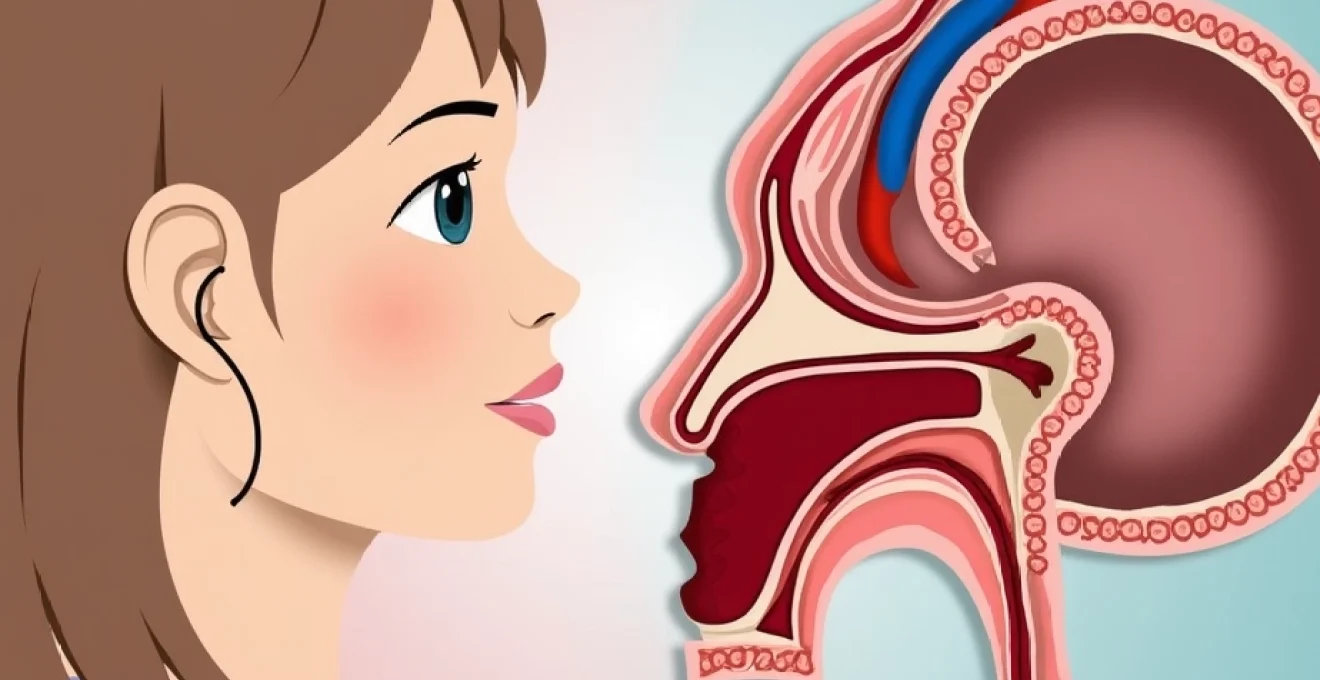
Anatomical changes in temporal bone and middle ear during pregnancy
Pregnancy induces subtle but significant anatomical changes throughout the body, including modifications to the temporal bone and middle ear structures that can contribute to pulsatile tinnitus development. These changes result from hormonal influences, increased blood volume, and altered tissue properties that affect sound transmission and perception. Understanding these anatomical adaptations helps explain why pulsatile tinnitus often emerges during specific stages of pregnancy and typically resolves postpartum.
The temporal bone houses critical auditory structures and serves as a conduit for major blood vessels, including the internal carotid artery and internal jugular vein. During pregnancy, hormonal influences cause generalised tissue swelling and increased vascular engorgement throughout the head and neck region. Temporal bone structures become more vascular and less acoustically dampening, creating conditions where blood flow sounds that were previously inaudible become clearly perceptible to the pregnant woman.
Oestrogen and progesterone particularly affect the mucosa lining the middle ear space and Eustachian tubes. This mucosal tissue becomes thickened and more vascular during pregnancy, altering the acoustic properties of the middle ear environment. The increased tissue mass and vascularity can act as a sound conductor rather than an insulator, facilitating transmission of vascular sounds from nearby blood vessels. Additionally, the softening effects of relaxin on connective tissues within the temporal bone may reduce the natural dampening that normally prevents internal sounds from reaching consciousness.
The middle ear’s three tiny bones – the malleus, incus, and stapes – along with their associated muscles and ligaments, undergo subtle changes during pregnancy. Hormonal influences can affect the tension and responsiveness of the stapedius and tensor tympani muscles, potentially altering their ability to modulate sound transmission. These microscopic changes, whilst individually minor, collectively create conditions that favour the perception of internal sounds, including the rhythmic pulsations of nearby blood vessels that characterise pulsatile tinnitus during pregnancy .
Differential diagnosis between benign pregnancy-related and pathological pulsatile tinnitus
Distinguishing between physiological pregnancy-related pulsatile tinnitus and symptoms indicative of serious pathology represents a critical clinical challenge. Benign pregnancy-related pulsatile tinnitus typically exhibits specific characteristics that differentiate it from pathological causes requiring immediate intervention. Understanding these distinguishing features enables healthcare providers to appropriately triage patients and avoid unnecessary anxiety whilst ensuring timely treatment of serious conditions.
Physiological pulsatile tinnitus during pregnancy usually develops gradually during the second trimester, coinciding with peak cardiovascular adaptations. The sounds are typically described as soft whooshing or rhythmic pulsing that matches the heart rate exactly. These benign symptoms often improve with position changes, particularly when the woman sits upright or lies on her left side to optimise venous return. Pregnancy-related physiological tinnitus rarely interferes with sleep or daily activities and is not associated with other neurological symptoms such as visual changes, severe headaches, or focal weakness.
In contrast, pathological pulsatile tinnitus often presents with alarming associated features that demand immediate medical attention. Warning signs include sudden onset of loud, intrusive pulsatile sounds that interfere with concentration or sleep, particularly when accompanied by severe headaches, visual disturbances, or blood pressure elevation above 140/90 mmHg. Pathological pulsatile tinnitus may present as unilateral symptoms that don’t improve with position changes and may actually worsen with certain head positions or activities that increase intracranial pressure.
The key to safe management lies in recognising red flag symptoms that distinguish dangerous pathology from benign pregnancy-related changes.
Temporal patterns provide valuable diagnostic clues in differentiating between benign and pathological causes. Physiological pregnancy-related pulsatile tinnitus tends to fluctuate throughout the day, often being most noticeable during quiet periods or when lying down. The intensity typically correlates with physical activity levels and may temporarily disappear during periods of distraction or background noise. Conversely, pathological causes often produce constant, unchanging symptoms that persist regardless of environmental factors or body position, creating a sense of relentless intrusion that significantly impacts quality of life.
Third trimester risk factors and postpartum pulsatile tinnitus resolution patterns
The third trimester presents unique challenges for pulsatile tinnitus management due to peak physiological stress on maternal cardiovascular and neurological systems. During this period, blood volume reaches maximum expansion, cardiac output peaks, and positional effects become more pronounced due to uterine size and foetal weight. Understanding third trimester risk factors enables healthcare providers to anticipate potential complications and implement appropriate monitoring strategies.
Several factors specifically increase pulsatile tinnitus risk during the third trimester. Maternal weight gain exceeding recommended guidelines can exacerbate intracranial pressure elevation, particularly in women predisposed to idiopathic intracranial hypertension. Multiple pregnancies place additional cardiovascular demands that can amplify vascular sounds, whilst advanced maternal age (over 35 years) increases the likelihood of developing pregnancy-induced hypertension. Third trimester complications such as gestational diabetes can also contribute to cardiovascular changes that worsen pulsatile tinnitus symptoms.
Positional factors become increasingly significant as pregnancy progresses into the third trimester. The enlarged uterus can compress major venous structures when the woman lies supine, forcing blood through collateral pathways that may include vessels near the temporal bone. This phenomenon, known as supine hypotensive syndrome, can dramatically worsen pulsatile tinnitus symptoms and explains why many women find relief when sleeping with elevated head positioning or lying on their left side to optimise venous return.
Sleep disturbances during the third trimester can create a vicious cycle where pulsatile tinnitus interferes with rest, leading to increased stress and blood pressure elevation that further exacerbates symptoms. The combination of physical discomfort from advanced pregnancy, anxiety about impending delivery, and persistent auditory symptoms can significantly impact maternal well-being during this critical period.
Most pregnancy-related pulsatile tinnitus follows predictable resolution patterns following delivery. The majority of women experience significant symptom improvement within the first week postpartum as blood volume begins normalising and hormonal levels start returning to baseline. Complete resolution typically occurs within 6-12 weeks postpartum, corresponding with the timeline for cardiovascular system recovery to pre-pregnancy state.
However, certain factors can delay or prevent complete resolution of symptoms. Women who developed pregnancy-induced hypertension may experience prolonged symptoms if blood pressure remains elevated postpartum. Breastfeeding mothers may notice fluctuating symptoms due to ongoing hormonal influences and increased metabolic demands. Iron deficiency anaemia, common in the postpartum period, can perpetuate symptoms by maintaining the hyperdynamic circulatory state that contributes to audible vascular sounds.
Persistent pulsatile tinnitus beyond 12 weeks postpartum warrants thorough evaluation to exclude underlying pathology that may have been masked by pregnancy-related changes. Conditions such as arteriovenous malformations, dural arteriovenous fistulas, or sigmoid sinus dehiscence may become apparent only after normal pregnancy-related cardiovascular adaptations have resolved. Early postpartum follow-up should include blood pressure monitoring, assessment for signs of postpartum pre-eclampsia, and evaluation of iron status to address potentially reversible causes of ongoing symptoms.
Understanding normal resolution patterns helps distinguish between expected postpartum recovery and pathological conditions requiring further investigation.
The psychological impact of persistent pulsatile tinnitus during the vulnerable postpartum period cannot be understated. New mothers already face significant challenges adjusting to parenthood, sleep deprivation, and physical recovery from childbirth. Ongoing auditory symptoms can contribute to anxiety, depression, and bonding difficulties that affect both maternal well-being and infant care. Healthcare providers should maintain awareness of these psychological dimensions whilst pursuing appropriate medical evaluation for persistent symptoms, ensuring comprehensive care that addresses both physical and emotional needs during this critical transition period.