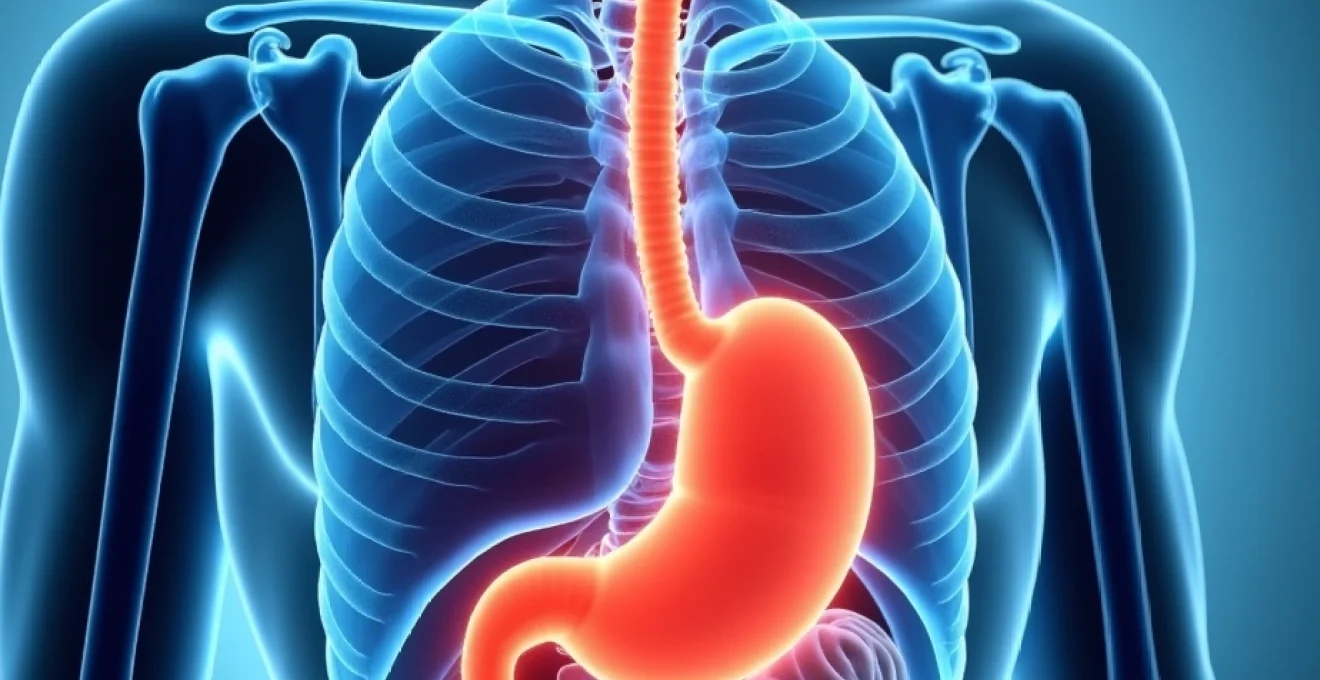
Hiatal hernias affect approximately 60% of individuals over the age of 60, yet many patients remain undiagnosed for years due to the condition’s propensity for manifesting through atypical symptoms. While gastroesophageal reflux disease represents the most recognised presentation, these anatomical abnormalities frequently produce a constellation of seemingly unrelated symptoms that can perplex both patients and clinicians. The displacement of gastric contents through the diaphragmatic hiatus creates a cascade of physiological disruptions that extend far beyond the digestive system, affecting cardiovascular, respiratory, and musculoskeletal function in ways that challenge conventional diagnostic approaches.
Atypical hiatal hernia manifestations beyond classic gastroesophageal symptoms
The complexity of hiatal hernia presentations stems from the intricate anatomical relationships between the stomach, diaphragm, and surrounding structures. When gastric tissue herniates through the esophageal hiatus, it creates mechanical compression and irritation that can generate symptoms in distant anatomical regions through shared neural pathways and referred pain mechanisms. Understanding these atypical presentations requires a comprehensive appreciation of how diaphragmatic displacement affects multiple organ systems simultaneously.
Research indicates that approximately 40% of patients with confirmed hiatal hernias experience symptoms that do not directly correlate with gastroesophageal dysfunction. These manifestations often include musculoskeletal pain patterns, particularly in the thoracic and cervical regions, which can lead to misdiagnosis and inappropriate treatment strategies. The temporal relationship between symptom onset and postural changes or food intake provides crucial diagnostic clues that distinguish hiatal hernia-related discomfort from primary musculoskeletal disorders.
Dorsal and intercostal pain patterns in type I sliding hiatal hernias
Type I sliding hiatal hernias frequently generate distinctive pain patterns along the dorsal spine, particularly affecting the mid-thoracic vertebrae between T6 and T10. This discomfort typically manifests as a dull, persistent ache that intensifies following meals or when assuming recumbent positions. The mechanism underlying this symptom involves mechanical irritation of the diaphragmatic crura and subsequent referred pain through the intercostal nerve distribution. Patients often describe the sensation as pressure-like discomfort that radiates horizontally along the rib cage, creating confusion with musculoskeletal conditions or even cardiac pathology.
Referred pain mechanisms through vagal and phrenic nerve pathways
The complex innervation of the diaphragm and gastroesophageal junction creates multiple pathways for referred pain transmission in hiatal hernia patients. The phrenic nerve, originating from cervical segments C3-C5, provides sensory innervation to the diaphragmatic pleura and can transmit pain signals to the shoulder and neck regions. Simultaneously, vagal afferent fibres carry visceral sensory information from the herniated gastric tissue, creating convergence points in the central nervous system that result in spatially distant pain perception . This neurological phenomenon explains why patients may experience shoulder blade pain or cervical discomfort without obvious local pathology.
Cervical and thoracic spine pain distribution in paraesophageal hernias
Paraesophageal hernias, representing Types II-IV hiatal hernias, demonstrate more pronounced effects on spinal pain patterns due to their larger size and greater mechanical impact. The herniated gastric fundus can create significant pressure against the posterior mediastinum, affecting the sympathetic chain and creating referred pain throughout the thoracic spine. Patients frequently report morning stiffness and progressive discomfort throughout the day, particularly after large meals when gastric distension exacerbates the herniation. The pain distribution often follows dermatome patterns, with T4-T8 segments most commonly affected, creating diagnostic challenges when cardiac pathology must be excluded.
Diaphragmatic Irritation-Induced scapular and shoulder blade discomfort
Direct irritation of the diaphragmatic surface by herniated gastric contents produces characteristic scapular pain that patients often describe as sharp, stabbing sensations beneath the shoulder blades. This symptom typically correlates with the degree of diaphragmatic elevation and can be reproduced through specific postural manoeuvres or deep inspiration. The phrenic nerve’s cervical origin explains why diaphragmatic irritation manifests as referred pain in the C4 dermatome distribution, encompassing the superior aspect of the shoulder and extending towards the neck. Understanding this referred pain pattern prevents unnecessary investigations for primary shoulder pathology while guiding clinicians towards appropriate diagnostic imaging of the thoracic cavity.
Cardiopulmonary symptoms masquerading as primary cardiac or respiratory conditions
Large hiatal hernias can produce a remarkable array of cardiopulmonary symptoms that frequently lead to extensive cardiac and respiratory evaluations before the correct diagnosis becomes apparent. The anatomical proximity of the herniated stomach to the heart and lungs creates mechanical compression effects that can significantly impact both cardiovascular and respiratory function. Emergency departments report that approximately 15-20% of patients presenting with chest pain and negative cardiac workups are subsequently found to have significant hiatal hernias contributing to their symptoms.
The diagnostic challenge intensifies when considering that hiatal hernia patients often belong to demographic groups at high risk for primary cardiac disease. Elderly patients, in particular, may undergo multiple cardiac catheterisations and stress tests before clinicians consider gastrointestinal causes for their chest discomfort. The overlap in symptom presentation necessitates a systematic approach to differential diagnosis that considers both the temporal relationship of symptoms to meals and their positional variation.
Exercise-induced dyspnoea and postural orthopnoea in large hiatal hernias
Massive hiatal hernias can significantly compromise respiratory function through direct compression of the lung bases and restriction of diaphragmatic excursion. Patients typically report progressive dyspnoea during physical exertion, which may be misattributed to primary pulmonary or cardiac pathology. The mechanism involves reduced functional residual capacity and impaired ventilation-perfusion matching in the compressed lung segments. Postural orthopnoea represents a particularly telling symptom, as the supine position allows greater gastric migration into the thoracic cavity, further compromising respiratory mechanics. Pulmonary function testing often reveals restrictive patterns with reduced total lung capacity and forced vital capacity measurements.
Atrial fibrillation and supraventricular tachycardia secondary to gastric distension
The phenomenon of gastrocardiac syndrome, though historically controversial, demonstrates clear mechanistic pathways in patients with large hiatal hernias. Gastric distension within the thoracic cavity can trigger cardiac arrhythmias through direct mechanical pressure on the left atrium and stimulation of cardiac autonomic reflexes. Studies indicate that up to 30% of patients with massive hiatal hernias experience episodic atrial fibrillation or supraventricular tachycardia, particularly following large meals. The temporal relationship between food intake, gastric distension, and arrhythmia onset provides crucial diagnostic evidence. Continuous cardiac monitoring during controlled feeding studies has documented reproducible arrhythmia induction in susceptible patients, confirming the mechanistic relationship between gastric herniation and cardiac electrical instability.
Pseudo-angina pectoris and Non-ST elevation myocardial infarction mimicry
Perhaps the most concerning presentation involves hiatal hernia patients who develop chest pain syndromes indistinguishable from acute coronary syndromes. The pain typically exhibits retrosternal location, pressure-like quality, and radiation to the left arm or jaw, fulfilling classical anginal criteria. However, careful history-taking often reveals postprandial exacerbation and positional variation that distinguishes hiatal hernia pain from true cardiac ischaemia. Electrocardiographic changes, including T-wave inversions and ST-segment depressions, can occur secondary to mechanical cardiac compression, further complicating the diagnostic process. Emergency physicians must maintain high clinical suspicion for hiatal hernia in patients with atypical coronary syndrome presentations , particularly when cardiac biomarkers remain normal despite suggestive symptomatology.
Compression asthma and restrictive lung pattern abnormalities
Large paraesophageal hernias can induce bronchospasm through mechanical compression of the lower respiratory tract and vagally mediated bronchial smooth muscle contraction. This compression asthma typically presents with wheeze and dyspnoea that correlates with gastric distension rather than traditional asthma triggers. Spirometry demonstrates reversible airway obstruction that improves with gastric decompression or positional changes that reduce the degree of herniation. The condition represents a form of extrinsic asthma that responds poorly to conventional bronchodilator therapy but shows dramatic improvement following surgical hernia repair. Recognition of this mechanism prevents inappropriate escalation of asthma medications while directing attention towards the underlying anatomical abnormality.
Neurological and systemic presentations of advanced hiatal hernia disease
Advanced hiatal hernia disease can produce a spectrum of neurological and systemic symptoms that extend far beyond the gastrointestinal tract. These presentations often develop insidiously over years, making recognition challenging for both patients and healthcare providers. The complex interplay between mechanical compression, autonomic dysfunction, and chronic inflammation creates a multisystem disorder that can significantly impact quality of life and functional capacity.
The prevalence of neurological symptoms in hiatal hernia patients approaches 25-30% in large case series, with symptoms ranging from subtle cognitive impairment to dramatic syncopal episodes. Understanding these diverse manifestations requires appreciation of how chronic gastric displacement affects multiple physiological systems simultaneously, creating cascading effects that can persist even after anatomical correction.
Vasovagal syncope episodes following gastric volvulus events
Gastric volvulus represents one of the most serious complications of paraesophageal hiatal hernias and can trigger profound vasovagal responses leading to syncopal episodes. The sudden gastric rotation creates intense visceral pain and triggers massive vagal stimulation, resulting in bradycardia and hypotension sufficient to cause loss of consciousness. These episodes typically occur following large meals when gastric distension predisposes to volvulus formation. Patients often describe prodromal symptoms including nausea, diaphoresis, and severe epigastric pain before losing consciousness. The temporal relationship to eating provides crucial diagnostic information that distinguishes gastric volvulus-induced syncope from primary cardiac or neurological causes of consciousness loss.
Chronic fatigue syndrome and Iron-Deficiency anaemia from occult bleeding
Chronic gastric irritation and mucosal erosion within the herniated gastric pouch can lead to occult gastrointestinal bleeding and subsequent iron-deficiency anaemia. This process typically develops gradually over months to years, creating a clinical picture resembling chronic fatigue syndrome. Cameron erosions, linear mucosal ulcerations occurring at the neck of large hiatal hernias, represent the primary source of chronic blood loss. Studies indicate that up to 40% of patients with large hiatal hernias develop some degree of iron deficiency, with 15-20% progressing to symptomatic anaemia. The fatigue associated with chronic anaemia often overshadows other hiatal hernia symptoms, leading to delayed diagnosis and inappropriate treatment with haematinics rather than definitive anatomical correction .
Sleep apnoea exacerbation through diaphragmatic displacement mechanisms
Large hiatal hernias can significantly worsen existing sleep apnoea or precipitate sleep-disordered breathing in previously asymptomatic individuals. The mechanism involves upward displacement of the gastric fundus, which reduces functional diaphragmatic excursion and alters respiratory mechanics during sleep. Additionally, gastroesophageal reflux associated with hiatal hernias can cause laryngeal oedema and upper airway inflammation, further compromising breathing during sleep. Polysomnography studies demonstrate that patients with large hiatal hernias exhibit more frequent apnoeic episodes and greater oxygen desaturation compared to controls. The relationship becomes particularly pronounced in the supine position, where gravitational effects maximise gastric migration into the thoracic cavity. Recognition of this association ensures that sleep study interpretation considers anatomical factors beyond traditional airway pathology.
Postural hypotension and autonomic dysfunction in elderly patients
Elderly patients with chronic hiatal hernias frequently develop subtle autonomic dysfunction manifesting as postural hypotension and impaired cardiovascular reflexes. The chronic vagal irritation from gastric herniation appears to disrupt normal autonomic balance, reducing the effectiveness of cardiovascular compensation mechanisms. This dysfunction becomes particularly apparent during positional changes, when normal baroreceptor responses fail to maintain adequate blood pressure. The condition often coexists with age-related autonomic decline, creating additive effects that significantly impact functional capacity. Tilt-table testing in affected patients demonstrates blunted heart rate responses and inadequate peripheral vasoconstriction during orthostatic stress. Understanding this relationship prevents attribution of symptoms to normal ageing processes while identifying patients who may benefit from surgical intervention to restore autonomic balance.
Diagnostic imaging protocols for identifying unusual hiatal hernia presentations
Accurate diagnosis of hiatal hernias presenting with atypical symptoms requires systematic imaging approaches that can demonstrate both anatomical abnormalities and functional consequences. Traditional imaging protocols designed for gastroesophageal reflux disease may inadequately characterise the complex three-dimensional relationships involved in large hiatal hernias, particularly when symptoms suggest cardiovascular or respiratory involvement. Modern imaging techniques must provide comprehensive evaluation of not only the hernia itself but also its impact on surrounding structures and organ systems.
The diagnostic challenge increases significantly when patients present with predominantly extragastrointestinal symptoms, as initial imaging studies may focus on the apparent organ system of concern rather than considering hiatal hernia as the underlying cause. Contrast-enhanced computed tomography has emerged as the gold standard for evaluating complex hiatal hernias, providing detailed anatomical information while allowing assessment of potential complications such as gastric volvulus or ischaemia.
Advanced imaging techniques have revolutionised the diagnosis of atypical hiatal hernia presentations, revealing the complex anatomical relationships that explain seemingly unrelated symptom patterns.
Barium studies remain valuable for functional assessment, particularly when evaluating swallowing mechanics and gastric emptying patterns. However, static radiographic images may underestimate the degree of herniation, as gastric contents can reduce spontaneously between imaging sequences. Dynamic studies performed with the patient in various positions provide more comprehensive information about the reducibility of the hernia and its relationship to symptom generation. The integration of multiple imaging modalities creates a comprehensive picture that guides both diagnostic and therapeutic decision-making.
Magnetic resonance imaging offers particular advantages in evaluating soft tissue relationships and can provide detailed assessment of diaphragmatic anatomy without radiation exposure. However, the technique requires longer acquisition times and may not adequately capture dynamic relationships during swallowing or position changes. The choice of imaging modality should consider both the clinical presentation and the specific anatomical information required for treatment planning. Multiplanar reconstructions allow visualisation of complex three-dimensional relationships that may not be apparent on conventional axial images.
Differential diagnosis considerations for atypical hiatal hernia symptoms
The diverse symptom complex associated with hiatal hernias creates significant diagnostic challenges, as many presentations overlap with common medical conditions affecting middle-aged and elderly populations. Systematic differential diagnosis requires consideration of multiple organ systems while maintaining awareness of the temporal relationships and positional variations that characterise hiatal hernia symptoms. The key diagnostic principle involves recognising symptom patterns that suggest mechanical compression or referred pain rather than primary pathology in the affected organ system.
Cardiovascular differential diagnosis represents perhaps the most critical consideration, given the potential for life-threatening conditions that require immediate intervention. The challenge intensifies when hiatal hernia patients develop genuine cardiac pathology, creating compound presentations that require careful clinical correlation. Serial cardiac biomarkers and electrocardiographic monitoring help distinguish true cardiac events from hiatal hernia-related chest pain, while the response to positional changes or antacid therapy provides additional diagnostic information.
The temporal relationship between symptoms and meals, combined with positional variation, provides the most reliable clinical indicators for distinguishing hiatal hernia symptoms from primary cardiac, respiratory, or musculoskeletal pathology.
Respiratory conditions, particularly in patients with underlying chronic obstructive pulmonary disease, require careful evaluation to determine the relative contributions of primary lung pathology versus mechanical compression from hiatal herniation. Pulmonary function testing before and after treatment can help quantify the functional impact of the
hernia and allows objective assessment of improvement following surgical intervention.
Musculoskeletal conditions frequently enter the differential diagnosis, particularly when patients present with thoracic spine pain or scapular discomfort. The distinction relies heavily on the relationship between symptoms and gastric distension, as primary musculoskeletal pathology typically demonstrates consistent pain patterns regardless of meal timing. Physical examination findings such as trigger points, range of motion limitations, and response to musculoskeletal interventions help clarify the diagnosis. However, clinicians must remain vigilant for concurrent conditions, as chronic pain patterns from hiatal hernia may lead to secondary musculoskeletal dysfunction through compensatory postural changes.
Neurological differential diagnosis becomes particularly complex when patients present with syncope, autonomic dysfunction, or sleep disturbances. Primary neurological conditions such as epilepsy, cardiac arrhythmias, or sleep apnoea may coexist with hiatal hernia, requiring careful evaluation of temporal relationships and response to treatment. The key diagnostic feature involves demonstrating improvement in neurological symptoms following gastric decompression or dietary modifications that reduce herniation. Advanced diagnostic techniques, including tilt-table testing and polysomnography, can help quantify the functional impact of hiatal hernia on autonomic and sleep physiology, providing objective evidence for the relationship between anatomical abnormality and neurological symptoms.
Successful diagnosis of atypical hiatal hernia presentations requires a high index of suspicion combined with systematic evaluation of symptom triggers, temporal patterns, and response to positional or dietary interventions.
The integration of clinical presentation, imaging findings, and response to therapeutic interventions provides the most reliable approach to diagnosing hiatal hernia in patients with atypical symptoms. This comprehensive evaluation strategy prevents both underdiagnosis of significant hiatal hernias and overattribution of symptoms to gastroesophageal pathology when other conditions may be responsible. The complexity of these diagnostic challenges underscores the importance of multidisciplinary collaboration between gastroenterologists, surgeons, cardiologists, and other specialists to ensure optimal patient outcomes.
Understanding the full spectrum of hiatal hernia presentations empowers healthcare providers to recognize this common yet frequently misdiagnosed condition across diverse clinical settings. Early identification and appropriate management of atypical presentations can significantly improve patient quality of life while preventing unnecessary investigations and treatments for presumed primary organ system pathology. The key to successful diagnosis lies in maintaining clinical awareness of the diverse manifestations possible with this anatomically complex condition, combined with systematic evaluation approaches that consider both typical and atypical symptom patterns.