When reviewing your medical imaging reports, you might encounter the phrase “spinal canal is patent” and wonder what this technical term means for your health. A patent spinal canal refers to an open, unobstructed passageway within your vertebral column where the spinal cord and cerebrospinal fluid flow freely. This finding typically indicates that there are no significant compressions, blockages, or structural abnormalities affecting the neural pathway through your spine. Understanding this terminology becomes particularly important when dealing with back pain, neurological symptoms, or following up on spinal conditions, as the patency status directly correlates with your spinal cord’s ability to function normally.
Understanding spinal canal anatomy and patent status definition
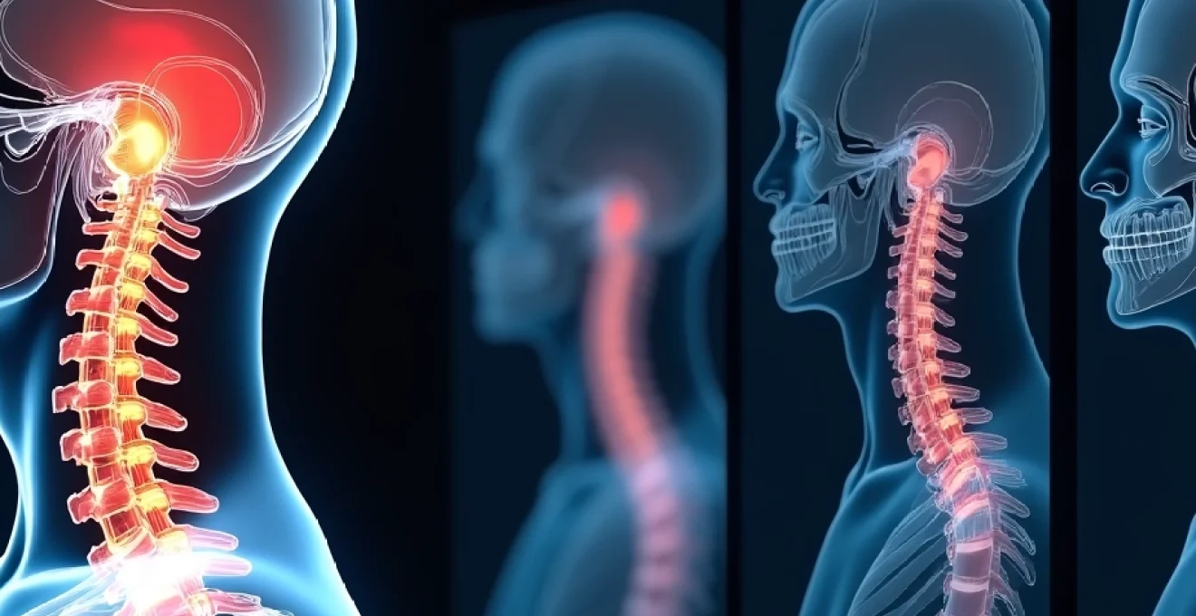
Anatomical structure of the vertebral canal from C1 to S5
The spinal canal extends from the first cervical vertebra (C1) at the base of your skull down to the fifth sacral vertebra (S5), forming a continuous bony tunnel that houses your spinal cord and related structures. This canal’s dimensions vary considerably throughout its length, with the cervical region measuring approximately 17-18mm in anteroposterior diameter, the thoracic region narrowing to about 15-16mm, and the lumbar region expanding again to 20-23mm. The canal’s triangular shape in the cervical spine transitions to a more circular configuration in the thoracic region, then becomes oval-shaped in the lumbar area.
Within this protective tunnel, the spinal cord occupies roughly 50-60% of the available space under normal conditions. The remaining space accommodates cerebrospinal fluid, blood vessels, nerve roots, and supportive tissues like the dura mater and ligamentum flavum. The canal’s posterior boundary consists of the laminae and ligamentum flavum , while the vertebral bodies and intervertebral discs form the anterior wall. Understanding these anatomical relationships helps explain why certain pathological conditions can compromise canal patency.
Cerebrospinal fluid flow dynamics within the thecal sac
Cerebrospinal fluid circulation within the patent spinal canal follows a complex pattern that supports spinal cord nutrition and waste removal. The thecal sac, which contains this fluid, extends from the foramen magnum to approximately the S2 level, creating a closed system where fluid flows both cephalad and caudad with cardiac and respiratory cycles. When the spinal canal maintains its patent status, this fluid can circulate freely, maintaining optimal pressure gradients and supporting normal neurological function.
Disruption of CSF flow patterns often serves as an early indicator of developing spinal stenosis or other pathological conditions. Modern imaging techniques can detect subtle changes in fluid dynamics even before structural compression becomes apparent on standard scans. This dynamic assessment proves particularly valuable when evaluating patients with intermittent neurological symptoms that may not correlate with static imaging findings.
Normal spinal cord dimensions and Cross-Sectional measurements
The spinal cord itself maintains relatively consistent dimensions throughout its length, though it does feature two natural enlargements corresponding to nerve supply for the upper and lower extremities. The cervical enlargement spans from C4 to T1 levels, while the lumbar enlargement extends from T9 to T12. In a patent canal, the cord’s transverse diameter measures approximately 13-14mm in the cervical region and 8-9mm in the thoracic area.
Clinical assessment of cord compression often relies on calculating the compression ratio, which compares the cord’s actual dimensions to expected normal values. A compression ratio exceeding 40% typically indicates significant pathological compression , even when the canal technically remains patent. This measurement becomes crucial when determining treatment approaches for conditions like cervical spondylotic myelopathy or thoracic disc herniation.
Patent vs stenotic canal: clinical distinction criteria
Distinguishing between a patent and stenotic spinal canal involves both quantitative measurements and qualitative assessment of tissue relationships. A patent canal maintains adequate space for neural structures without causing compression symptoms, while stenosis implies narrowing that may or may not produce clinical manifestations. The transition from patent to stenotic status often occurs gradually, making early detection challenging without systematic evaluation protocols.
Clinical stenosis requires not just anatomical narrowing but also the presence of neurological symptoms or signs that correlate with the imaging findings.
MRI and CT imaging protocols for spinal canal assessment
T1-weighted and T2-Weighted sequence interpretation
T1-weighted MRI sequences excel at demonstrating anatomical detail and tissue contrast within the spinal canal, particularly for identifying fat planes and distinguishing between different soft tissue structures. In a patent canal, you’ll observe clear cerebrospinal fluid signal surrounding the spinal cord, with well-defined borders between neural tissue and adjacent structures. The normal spinal cord appears as intermediate signal intensity on T1-weighted images, while CSF demonstrates low signal intensity, creating excellent contrast for assessing canal patency.
T2-weighted sequences provide superior visualization of pathological processes and fluid accumulation within the spinal canal. A patent spinal canal on T2-weighted imaging shows bright CSF signal completely surrounding the spinal cord , without areas of signal loss or compression. These sequences prove particularly valuable for detecting early myelomalacia, syrinx formation, or subtle cord compression that might not be apparent on T1-weighted images alone.
Sagittal and axial plane analysis techniques
Sagittal plane imaging provides an excellent overview of spinal canal patency throughout its entire length, allowing radiologists to identify areas of narrowing, assess alignment, and evaluate the relationship between spinal cord and canal dimensions. The normal sagittal appearance shows continuous CSF signal both anterior and posterior to the spinal cord, with consistent canal dimensions that follow predictable anatomical variations.
Axial plane analysis becomes essential for quantifying the degree of canal compromise and understanding the spatial relationships between compressive structures and neural elements. Cross-sectional measurements obtained from axial images provide the most accurate assessment of residual canal diameter and help distinguish between central stenosis, lateral recess stenosis, and foraminal narrowing. This multi-planar approach ensures comprehensive evaluation of canal patency status.
Contrast enhancement patterns in myelographic studies
CT myelography remains valuable for assessing spinal canal patency, particularly when MRI is contraindicated or when dynamic evaluation is necessary. Intrathecal contrast agent distribution patterns provide direct visualization of CSF spaces and can reveal subtle stenosis not apparent on non-contrast studies. In a patent canal, contrast flows freely throughout the thecal sac, creating smooth, continuous columns of enhancement around neural structures.
Contrast pooling, abrupt cutoffs, or irregular flow patterns may indicate areas of compromise even when static images suggest adequate canal dimensions. Dynamic myelographic studies can reveal positional stenosis that only becomes apparent with flexion or extension maneuvers, providing valuable information for surgical planning and patient counseling.
Quantitative measurements using Torg-Pavlov ratio
The Torg-Pavlov ratio represents a standardized method for assessing cervical spinal canal dimensions, calculated by dividing the canal diameter by the vertebral body width on lateral radiographs. Values below 0.80 suggest developmental stenosis, while ratios above 1.00 typically indicate a patent canal with adequate reserve capacity. This measurement proves particularly useful for screening athletes and evaluating patients with cervical spine trauma.
Modern imaging software allows for precise digital measurements that enhance the accuracy of Torg-Pavlov ratio calculations compared to traditional manual methods. These quantitative assessments complement qualitative imaging interpretation and provide objective criteria for determining canal patency status. However, clinicians must remember that anatomical measurements don’t always correlate directly with functional capacity or symptom severity.
Clinical conditions associated with Non-Patent spinal canals
Central spinal stenosis pathophysiology and grading
Central spinal stenosis develops through various pathological processes that gradually compromise spinal canal patency, most commonly involving degenerative changes in the facet joints, ligamentum flavum hypertrophy, and disc bulging or herniation. These age-related changes typically progress slowly, allowing the nervous system to adapt until compensation mechanisms fail and symptoms emerge. The pathophysiology involves both mechanical compression and vascular compromise of neural structures.
Stenosis grading systems help standardize the assessment of canal compromise severity. Mild stenosis typically involves less than 30% canal narrowing, moderate stenosis encompasses 30-50% narrowing, while severe stenosis indicates greater than 50% canal compromise. These classifications guide treatment decisions and help predict patient outcomes following various therapeutic interventions. Understanding stenosis severity becomes crucial when counseling patients about prognosis and treatment options.
Ligamentum flavum hypertrophy impact on canal diameter
The ligamentum flavum normally measures 2-3mm in thickness but can hypertrophy to 5-10mm or more in degenerative conditions, significantly reducing spinal canal patency. This elastic ligament loses its normal properties with age, becoming fibrotic and less compliant, which contributes to posterior canal compromise. The hypertrophy process often occurs asymmetrically, creating irregular canal narrowing patterns that can be difficult to assess on imaging studies.
Ligamentum flavum hypertrophy frequently occurs in conjunction with facet joint arthropathy and may contribute to neurogenic claudication symptoms even when disc pathology appears minimal. Dynamic imaging studies can reveal the true extent of canal compromise caused by ligamentum flavum buckling during spinal extension, which may not be apparent on neutral position scans. This understanding influences surgical approaches and helps explain why some patients experience positional symptom variation.
Disc herniation effects on thecal sac compression
Intervertebral disc herniation can rapidly transform a patent spinal canal into a significantly compromised space, particularly when large central herniations occur in patients with pre-existing developmental or degenerative narrowing. The mechanism involves displacement of nucleus pulposus material beyond the normal confines of the annulus fibrosus, creating mass effect on neural structures within the canal.
The severity of thecal sac compression depends not only on herniation size but also on the baseline canal dimensions and the specific location of the displaced disc material. Patients with constitutionally narrow canals may develop severe symptoms from relatively small herniations that would be asymptomatic in individuals with generous canal dimensions. This relationship explains the wide variation in clinical presentations among patients with similar imaging findings.
Congenital spinal canal narrowing syndromes
Developmental spinal stenosis results from congenital variations in vertebral formation that create inherently narrow spinal canals, predisposing affected individuals to earlier symptom onset with degenerative changes. These conditions include achondroplasia, where shortened pedicles and thickened laminae reduce canal dimensions, and idiopathic developmental stenosis, which may affect single levels or multiple segments throughout the spine.
Patients with congenital canal narrowing often experience symptoms at younger ages and may require earlier surgical intervention compared to those with purely degenerative stenosis.
Radiological reporting standards for patent spinal canals
Standardized radiological reporting of spinal canal patency requires consistent terminology and systematic assessment protocols to ensure clear communication between imaging specialists and treating physicians. Reports should specify the exact levels evaluated, measurement techniques employed, and any limitations of the study that might affect interpretation accuracy. Comprehensive reporting includes assessment of both static and dynamic factors that influence canal patency, particularly when evaluating patients with position-dependent symptoms.
Modern reporting standards emphasize the importance of correlating imaging findings with clinical presentation and providing specific measurements when possible. Radiologists increasingly include quantitative data such as canal dimensions, cross-sectional areas, and compression ratios to support their qualitative assessments. This approach facilitates treatment planning and helps establish objective criteria for monitoring disease progression or treatment response over time.
Quality assurance measures in spinal imaging reporting include standardized measurement protocols, peer review processes, and regular calibration of measurement tools to ensure consistency across different observers and imaging systems. Structured reporting templates help ensure that all relevant aspects of canal patency are addressed while reducing the likelihood of omitting important findings that could influence patient management decisions.
Clinical implications of patent spinal canal findings
A patent spinal canal finding on imaging studies generally indicates that you have adequate space for neural structures and normal cerebrospinal fluid flow, which typically correlates with preserved neurological function and absence of compression-related symptoms. However, clinicians must interpret these findings within the broader clinical context, as some patients may experience symptoms despite apparently adequate canal dimensions on static imaging studies. The relationship between anatomical patency and functional capacity involves complex interactions between neural tissue, vascular supply, and biomechanical factors.
Patent canal findings can provide reassurance to patients with spine-related concerns, particularly when symptoms might be attributed to non-structural causes such as muscle tension, postural problems, or referred pain from other sources. Understanding that your spinal canal remains open and uncompromised can help reduce anxiety and guide treatment toward appropriate conservative management strategies rather than surgical interventions.
Long-term prognosis for patients with patent spinal canals generally remains favorable, though regular monitoring may be appropriate in cases where degenerative changes are evident but not yet causing significant compromise. Preventive measures such as maintaining proper posture, engaging in appropriate exercise programs, and managing risk factors for spinal degeneration become particularly important for preserving canal patency over time.
Differential diagnosis when spinal canal patency is questioned
When imaging studies reveal questionable spinal canal patency, clinicians must consider various conditions that can mimic or coexist with spinal stenosis. Peripheral arterial disease can produce claudication symptoms similar to neurogenic claudication, while hip arthritis or piriformis syndrome may cause referred pain patterns that resemble spinal pathology. Comprehensive clinical evaluation becomes essential for distinguishing between these conditions and determining the true source of patient symptoms.
Advanced imaging techniques such as dynamic MRI, upright imaging, or specialized sequences may be necessary when standard studies fail to explain symptom patterns in patients with apparently patent canals. These specialized studies can reveal positional stenosis, ligamentous instability, or subtle compression that becomes apparent only under physiological loading conditions. Understanding when to pursue additional imaging helps optimize diagnostic accuracy while avoiding unnecessary testing.
The concept of functional stenosis involves situations where canal dimensions appear adequate on imaging but neural function remains compromised due to factors such as inflammation, ischemia, or metabolic dysfunction. This phenomenon explains why some patients continue to experience neurological symptoms despite patent canal findings and highlights the importance of comprehensive neurological evaluation in addition to imaging assessment. Treatment approaches for functional stenosis may differ significantly from those used for mechanical compression, emphasizing the need for accurate differential diagnosis.