Hip discomfort when crossing your legs affects millions of individuals daily, yet many dismiss this pain as merely a minor inconvenience. This seemingly simple posture creates complex biomechanical changes within the hip joint that can trigger or exacerbate various musculoskeletal conditions. The act of crossing one leg over another places the hip in an extreme position of flexion, adduction, and internal rotation, fundamentally altering the normal mechanics of this ball-and-socket joint.
Understanding why this common sitting position causes pain requires examining the intricate relationship between hip anatomy, muscle function, and joint mechanics. Modern sedentary lifestyles have made crossed-leg positioning increasingly prevalent, contributing to a rise in hip-related complaints among office workers, students, and professionals who spend extended periods seated. The consequences extend far beyond temporary discomfort, potentially leading to chronic pain patterns and functional limitations that impact daily activities.
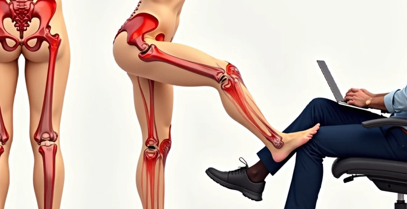
Anatomical structure of the hip joint and crossing leg mechanics
The hip joint represents one of the body’s most stable and powerful articulations, designed to support body weight while allowing multi-directional movement. This ball-and-socket configuration consists of the femoral head nestled within the acetabular socket of the pelvis, surrounded by a complex network of muscles, ligaments, and supportive structures. When functioning optimally, this arrangement permits smooth movement across multiple planes while maintaining structural integrity under significant loads.
Acetabular socket positioning during leg crossing movement
During leg crossing, the acetabular socket must accommodate extreme positioning of the femoral head, creating abnormal contact pressures within the joint. The acetabulum, naturally angled to provide optimal coverage of the femoral head in neutral positioning, becomes compromised when the leg moves into the crossed position. This altered relationship increases stress concentrations on specific areas of the joint cartilage, particularly the anterosuperior rim where impingement commonly occurs.
The acetabular labrum, a fibrocartilaginous rim that deepens the socket and enhances joint stability, experiences heightened tension during leg crossing. This structure, already vulnerable to wear and tear with age, faces additional stress as it attempts to maintain joint congruency in this non-physiological position. Repetitive stress on the labrum can lead to microscopic tears that eventually progress to symptomatic pathology.
Femoral head impingement against joint capsule
The femoral head’s position during leg crossing brings it into abnormal contact with the joint capsule, creating a pinching effect known as femoroacetabular impingement. This mechanical conflict occurs when the femoral neck contacts the acetabular rim, compressing soft tissues and restricting normal joint motion. The resulting compression can cause immediate discomfort and contribute to long-term degenerative changes within the joint.
Cam-type and pincer-type impingement patterns both become exaggerated during leg crossing, regardless of pre-existing hip morphology. Individuals with subtle anatomical variations that predispose them to impingement may experience their first symptoms when adopting crossed-leg positions regularly. The combination of hip flexion beyond 90 degrees with simultaneous adduction and internal rotation creates the perfect storm for impingement-related pain.
Piriformis muscle compression on sciatic nerve pathway
The piriformis muscle plays a crucial role in hip stability and external rotation, but crossing your legs places this muscle in a lengthened, stressed position. Located deep within the buttock region, the piriformis can compress the sciatic nerve when tight or inflamed, creating symptoms that radiate down the posterior thigh. This condition, known as piriformis syndrome, becomes more likely when individuals habitually cross their legs.
Sciatic nerve compression occurs because the nerve typically passes beneath or through the piriformis muscle belly. When the muscle becomes tight from prolonged stretching during leg crossing, it can create a tourniquet effect on the nerve. This compression results in numbness, tingling, or burning sensations that may be mistaken for hip joint pathology when the primary issue lies within the soft tissue structures.
Iliopsoas tendon stretch and tension distribution
The iliopsoas complex, consisting of the iliacus and psoas major muscles, functions as the primary hip flexor and plays a vital role in core stability. During leg crossing, these muscles experience simultaneous lengthening and rotational stress that can lead to tendon irritation and pain. The iliopsoas tendon passes over the femoral head and can become compressed against bony structures when the hip is positioned in extreme flexion with adduction.
Iliopsoas tendinopathy develops gradually through repetitive stress, often beginning as minor discomfort that progresses to significant pain with movement. The condition becomes particularly problematic for individuals who cross their legs frequently, as the sustained stretch weakens the muscle-tendon unit while simultaneously increasing tension on the structure. This paradoxical situation creates a cycle of dysfunction that perpetuates hip pain.
Common hip pathologies triggered by Crossed-Leg positioning
Crossed-leg positioning acts as a catalyst for various hip pathologies, either initiating new conditions or exacerbating existing ones. Research indicates that up to 90% of individuals experiencing lateral hip pain have conditions other than simple bursitis, with tendinopathy and muscle imbalances being predominant factors. The mechanical stress imposed by frequent leg crossing contributes significantly to these statistics, creating patterns of dysfunction that affect multiple anatomical structures simultaneously.
Femoroacetabular impingement syndrome exacerbation
Femoroacetabular impingement (FAI) represents a significant cause of hip pain in active individuals, with crossed-leg positioning serving as a primary aggravating factor. This condition involves abnormal contact between the femoral neck and acetabular rim, creating a pinching sensation that worsens with hip flexion and internal rotation. The prevalence of FAI has increased substantially in recent years, partly attributed to prolonged sitting postures and repetitive hip flexion activities.
Cam-type impingement occurs when an abnormal bump on the femoral neck contacts the acetabular cartilage, while pincer-type impingement results from over-coverage of the acetabulum. Both variants become symptomatic more readily when individuals regularly cross their legs, as this position maximises the impingement mechanism. The resulting cartilage damage and labral tears can progress to osteoarthritis if left untreated, making early recognition and intervention crucial.
Greater trochanteric bursitis inflammatory response
Greater trochanteric pain syndrome, commonly referred to as hip bursitis, affects approximately 20-25% of the population at some point in their lives. This condition involves inflammation of the trochanteric bursa, a fluid-filled sac that reduces friction between the greater trochanter and overlying soft tissues. Crossed-leg positioning places additional stress on this bursa through increased tension in the iliotibial band and gluteal tendons.
The inflammatory response associated with trochanteric bursitis creates a cycle of pain and dysfunction that becomes self-perpetuating.
Pain almost always goes away with targeted exercise and stretching, but avoiding aggravating positions like leg crossing remains essential for successful treatment.
Women experience this condition four times more frequently than men, particularly during and after menopause due to hormonal changes affecting soft tissue health.
Labral tear progression through repetitive stress
Acetabular labral tears represent a significant source of hip pain, particularly in younger, active individuals. The labrum serves multiple functions, including joint stability, shock absorption, and maintenance of joint fluid pressure. Crossed-leg positioning places abnormal stress on specific regions of the labrum, particularly the anterior and anterosuperior portions where tears most commonly occur.
Degenerative labral tears often begin as minor fraying but progress to full-thickness tears with continued mechanical stress. The avascular nature of much of the labral tissue means that healing potential remains limited, making prevention through proper positioning crucial. Individuals who frequently cross their legs may unknowingly accelerate labral degeneration, leading to chronic hip pain that requires surgical intervention.
Hip flexor tendinopathy development patterns
Hip flexor tendinopathy affects the tendons responsible for lifting the thigh toward the abdomen, primarily the iliopsoas and rectus femoris. This condition develops through repetitive stress and overuse, with crossed-leg positioning contributing significantly to the problem. The sustained stretch placed on hip flexors during leg crossing creates an eccentric loading pattern that can lead to tendon degeneration over time.
Tendinopathy development follows a predictable pattern, beginning with reactive tendon changes and potentially progressing to degenerative pathology. Early intervention focusing on load management and movement modification can prevent progression to chronic stages. However, individuals who continue crossing their legs despite early symptoms often experience worsening pain and functional limitations that require more intensive treatment approaches.
Biomechanical analysis of hip rotation during leg crossing
The biomechanics of leg crossing involve complex three-dimensional movements that challenge the hip joint’s normal function. When you cross your legs, the hip undergoes simultaneous flexion, adduction, and internal rotation – a combination that places maximum stress on joint structures. This triplanar movement pattern exceeds the hip’s optimal functional range, creating compensation strategies throughout the kinetic chain that can perpetuate pain and dysfunction.
Hip flexion during leg crossing typically exceeds 100 degrees, surpassing the normal functional range required for most daily activities. Combined with adduction angles approaching 30 degrees and internal rotation of 20-40 degrees, this positioning creates what biomechanists term a “close-packed” position where joint surfaces experience maximum contact pressure. The resulting stress concentration affects both articular cartilage and surrounding soft tissues, explaining why many individuals experience immediate discomfort when crossing their legs.
Internal rotation represents the most problematic component of this movement pattern, as the hip joint has limited natural capacity for this motion. Modern sedentary lifestyles often result in reduced internal rotation range of motion, making the crossed-leg position even more stressful for contemporary individuals. Range of motion deficits in internal rotation force compensatory movements in adjacent joints, creating a cascade of biomechanical dysfunction that extends beyond the hip joint itself.
The temporal aspects of leg crossing also significantly impact joint health. Brief periods of crossed-leg positioning may cause minimal harm, but sustained postures lasting 30 minutes or longer begin to produce measurable changes in muscle tension and joint pressure. Research demonstrates that prolonged crossed-leg sitting reduces blood flow to lower extremity muscles while simultaneously increasing compressive forces within the hip joint, creating an environment conducive to tissue damage and pain development.
Muscular compensation patterns and pain referral mechanisms
Crossed-leg positioning triggers complex muscular compensation patterns that extend far beyond the immediate hip region. The body’s attempt to maintain stability and comfort in this non-optimal position results in altered muscle activation patterns that can perpetuate dysfunction long after the legs are uncrossed. Understanding these compensation mechanisms provides crucial insight into why hip pain often persists despite avoiding the aggravating position.
Primary compensations begin with the deep hip rotators, particularly the piriformis, gemelli, and obturator muscles, which must work overtime to stabilise the femoral head in its altered position. These muscles become overactive and tight, leading to trigger point development and referral patterns that can mimic hip joint pathology. Simultaneously, the gluteal muscles, particularly the gluteus medius and minimus, become lengthened and weakened, losing their ability to provide adequate hip stability during functional activities.
Secondary compensations involve the lumbopelvic region, where the pelvis must accommodate the asymmetrical positioning of the crossed legs. The sacroiliac joint experiences altered loading patterns, while the lumbar spine adopts compensatory curves to maintain upright posture. These adaptations can create referred pain patterns that manifest as hip discomfort, complicating the diagnostic process and treatment planning.
The body isn’t always smart in recognising where the pain is coming from, and spine arthritis, a pinched nerve, or bones in the spine rubbing together can create pain in the side of your hip.
This phenomenon highlights the interconnected nature of the musculoskeletal system and emphasises the importance of comprehensive assessment when evaluating hip pain complaints.
Pain referral mechanisms also involve the nervous system’s response to sustained mechanical stress. Prolonged compression of neural structures during leg crossing can sensitise pain pathways, creating hyperalgesia and allodynia that persist beyond the initial mechanical irritation. This neurological component explains why some individuals continue experiencing hip pain even after modifying their sitting habits and addressing mechanical factors.
Diagnostic approaches for Cross-Legged hip pain assessment
Accurate diagnosis of cross-legged hip pain requires a systematic approach that considers both mechanical and neurological factors. Healthcare providers must differentiate between true hip joint pathology and referred pain from surrounding structures, as treatment strategies vary significantly based on the underlying cause. The diagnostic process typically begins with a thorough history that identifies specific aggravating factors, pain patterns, and functional limitations associated with crossed-leg positioning.
Physical examination plays a crucial role in identifying the source of hip pain, with specific tests designed to reproduce symptoms and assess joint function. The FADIR test (flexion, adduction, internal rotation) directly mimics the crossed-leg position and can help identify femoroacetabular impingement or labral pathology. Conversely, the FABER test (flexion, abduction, external rotation) assesses different aspects of hip function and can differentiate between intra-articular and extra-articular sources of pain.
Advanced imaging techniques provide valuable information when clinical examination findings remain unclear. Magnetic resonance imaging (MRI) offers detailed visualisation of soft tissue structures, including the labrum, cartilage, and surrounding muscles. MRI arthrography , involving intra-articular contrast injection, enhances the ability to detect labral tears and cartilage defects that might not be visible on conventional imaging. However, imaging findings must always be correlated with clinical presentation, as asymptomatic labral tears and cartilage changes are common in the general population.
Diagnostic injections serve both therapeutic and diagnostic purposes, helping to localise pain sources when multiple pathologies coexist. Intra-articular hip injections can differentiate between hip joint pathology and extra-articular causes of pain, while trochanteric bursa injections help identify greater trochanteric pain syndrome. The temporary relief provided by these injections confirms the diagnosis and guides subsequent treatment decisions.
Therapeutic interventions and movement modification strategies
Successful management of cross-legged hip pain requires a multi-faceted approach that addresses both immediate symptoms and underlying mechanical dysfunction. Conservative treatment remains the first-line approach for most conditions, with surgery reserved for cases that fail to respond to non-operative management. The key to successful treatment lies in identifying and modifying the specific factors that perpetuate pain while simultaneously addressing tissue dysfunction through targeted interventions.
Movement modification represents the cornerstone of treatment, beginning with education about proper sitting postures and alternative leg positions. Patients must understand the biomechanical rationale for avoiding crossed-leg positioning and learn practical strategies for maintaining comfort during prolonged sitting. Alternative positions include keeping both feet flat on the floor, using a footrest to maintain hip flexion at 90 degrees, or alternating between different sitting positions throughout the day.
Exercise therapy focuses on restoring muscle balance and joint function through targeted strengthening and stretching programs. Hip abductor strengthening, particularly of the gluteus medius and minimus, helps restore lateral hip stability and reduces compensatory patterns. Core strengthening exercises improve overall postural control and reduce the tendency to adopt problematic sitting positions. Flexibility exercises targeting the hip flexors, adductors, and external rotators help restore normal joint range of motion and reduce tissue tension.
Manual therapy techniques, including massage, joint mobilisation, and trigger point release, address soft tissue restrictions and joint stiffness that contribute to pain and dysfunction. These interventions work synergistically with exercise therapy to restore normal movement patterns and reduce pain sensitivity. The combination of manual therapy and exercise has proven more effective than either intervention alone for most hip pain conditions.
Advanced therapeutic options include injection therapies, such as corticosteroid injections for inflammatory conditions or platelet-rich plasma (PRP) for tendinopathy. These interventions can provide temporary symptom relief while allowing patients to engage more fully in rehabilitation programs. However, the success of injection therapy depends heavily on concurrent movement modification and exercise compliance, as failure to address underlying mechanical factors often results in symptom recurrence.